
Diversity and Antimicrobial Resistance Profile of Bacteria Isolated from Sheep Mastitis and Bulk-Tank Milk in the East-Centre Region of Portugal
M.Aires Pereira*, A. Lameira Baptista, M. Rosário, R. M. da Silva Cruz, F. A. de Almeida Esteves, J. Madanelo, C. L. de Vasconcelos Nóbrega, A. C. Pais Mega de Andrade, C. Garcia, C. Coelho, C. S. Arede dos Santos, M. Malva, H. Vala and T. Ferreira
Maria AIRES. PEREIRA1,2,3* (corresponding author), mapereira@esav.ipv.pt, orcid.org/0000-0002-4054-9124; Alexandra LAMEIRA BAPTISTA1,2,4, alexabaptista@esav.ipv.pt, orcid.org/0009-0002-8137-1139; Mariana ROSÁRIO1, mariana.rosario04@gmail.com, orcid.org/0009-0001-6429-3573; Rita Marisa da SILVA CRUZ 1,5,6, rcpaiva@esav.ipv.pt, orcid.org/0000-0002-5767-7835; Fernando Alexandre de ALMEIDA ESTEVES1,2 festeves@esav.ipv.pt, orcid.org/0000-0003-0589-0746; João MADANELO1; joao.madanelo@gmail.com; Carmen Lucia de VASCONCELOS NOBREGA1,7 cnobrega@esav.ipv.pt, orcid.org/0000-0003-3941-799X; Ana Cristina PAIS MEGA DE ANDRADE 1,2, amega@esav.ipv.pt, orcid.org/0000-0002-9913-875X; Carla GARCIA1, cgarcia@esav.ipv.pt; Catarina COELHO1,2, ccoelho@esav.ipv.pt, orcid/org0000-0002-5272-7303; Carla Sofia AREDE DOS SANTOS 1, casarede@esav.ipv.pt, orcid.org/0000-0001-5908-8500; Madalena MALVA8 malva@esav.ipv.pt, orcid.org/0000-0003-4982-0184; Helena VALA1,2,9 h.vala@sc.ipv.pt, orcid.org/0000-0001-6829-4867; Tiago FERREIRA10 tiagoandreferreir@gmail.com.
1Instituto Politécnico de Viseu, Escola Superior Agrária de Viseu, Av. Dr. António Almeida Henriques, 3500-631 Viseu, Portugal;
2CERNAS-IPV Research Centre, Instituto Politécnico de Viseu, Campus Politécnico, Repeses, 3504-510 Viseu, Portugal;
3Global Health and Tropical Medicine, GHTM, Associate Laboratory in Translation and Innovation Towards Global Health, LA-REAL, Instituto de Higiene e Medicina Tropical, IHMT, Universidade NOVA de Lisboa, UNL, Rua da Junqueira 100, 1349-008 Lisboa, Portugal;
4 Universidade de Trás-os-Montes e Alto Douro, Quinta de Prados 5000-801, Vila Real, Portugal;
5Epidemiology Research Unit (EPIUnit), Instituto de Saúde Pública da Universidade do Porto, Rua das Taipas 135, 4050-600, Porto, Portugal;
6Laboratory for Integrative and Translational Research in Population Health (ITR), Rua das Taipas 135, 4050-600, Porto, Portugal;
7Center for the Research and Technology of Agro-Environmental and Biological Sciences (CITAB), University of Trás-os-Montes e Alto Douro, Quinta de Prados, Edifício Reitoria, 5000-801, Vila Real, Portugal;
8Instituto Politécnico de Viseu, Escola Superior de Tecnologia e Gestão de Viseu, Avenida Cidade Politécnica, 3504-510, Viseu, Portugal;
9Veterinary and Animal Research Centre (CECAV), UTAD, Associate Laboratory for Animal and Veterinary Sciences (AL4AnimalS), Quinta de Prados, Apartado 1013, 5000-801, Vila Real, Portugal;
10Australian Laboratory Services (ALS), Zona Industrial de Tondela Lote 6, 3460-070, Tondela, Portugal.
![]()
https://doi.org/10.46419/cvj.57.5.9
Abstract
The microbiological quality of milk reflects both udder health and the subsequent contamination that can occur through direct and indirect reservoirs. This study aimed to identify bacterial diversity in milk samples from ewes with mastitis and in bulk-tank milk, and to investigate their antibiotic susceptibility/resistance profiles. A total of 232 milk samples were analysed and 22 bacterial species were isolated, with Staphylococcus aureus (20.3%) and S. epidermidis (18.6%) as the dominant species. The diversity of bacterial species isolated from the bulk-tank was higher and included 44 species, dominated by E. coli (20.6%), S. aureus (7.8%) and Hafnia alvei (7.2%). Antimicrobial susceptibility testing was performed on 217 bacterial isolates. E. coli isolated from individual milk samples revealed high rates of resistance to penicillins and tetracyclines, whereas S. aureus revealed high rates of resistance to aminoglycosides, polymyxin, sulfonamides and tetracycline, maintaining high levels of susceptibility to third generation cephalosporins, lincosamides, some macrolides, penicillins, phenicols and pleuromutilins. The resistance profile of bacteria isolated from the bulk-tank milk was more concerning than that of the milk of ewe with mastitis. These results point to the need to implement structural improvements on farms and raise awareness about strengthening biosecurity, medical prophylaxis and control of access to veterinary medicines to improve milk quality and reduce the use of antibiotics.
Keywords: Escherichia coli; coagulase-negative Staphylococcus; Staphylococcus aureus; intramammary infection; bulk-tank milk; antimicrobial susceptibility testing
Introduction
Cheese production is an important economic activity in the East-Centre region of Portugal, and follows artisanal procedures passed down through generations of shepherds (Macedo et al., 1993; Tavaria and Malcata, 2000; Reis Lima et al., 2019). Protected Designation of Origin (PDO) and Indication Protected Geographical (IGP) sheep cheese made of raw milk is highly prized by the Portuguese population, and considered one of the primary representative products of the Centre region of Portugal, with qualities and characteristics specific to this specific geographical area (Turismo de Portugal; Hilali et al., 2011).
The composition of milk microbiota has a direct impact on the quality of milk and dairy products. While some bacteria found in milk are technologically important as they are involved in flavour, aroma and colour development in cheese (Quigley et al., 2013), others may raise public health concerns, including Escherichia coli, Staphylococcus aureus, Enterococcus spp., Streptococcus spp., Listeria spp., Salmonella spp., Campylobacter spp., Clostridium perfrigens, among others (Almeida et al., 2007; Quigley et al., 2013; Praça et al., 2023; Iancu et al., 2024).
The health of the mammary gland is essential to produce high-quality milk. Bacterial intramammary infection (IMI) is one of the most costly and severe diseases in sheep dairy farms, negatively impacting milk yield and quality, as well as animal health and welfare (Contreras and Rodríguez, 2011). S. aureus and coagulase-negative Staphylococcus (CNS) are the main causes of clinical and subclinical IMI, respectively (Bergonier et al., 2003; Olechnowicz and Ja´skowski, 2014). The most frequently isolated CNS species are Staphylococcus epidermidis, S. simulans, S. xylosus, and S. chromogenes (Moroni et al., 2005; Vanderhaeghen et al., 2014).
Laboratory diagnosis, including culture-based and antimicrobial susceptibility testing (AST), is essential for the effective treatment of IMI (Iancu et al., 2024). On the other hand, bulk-tank milk analysis allows monitoring the hygienic quality of the milk, helping to identify poor management practices (Barrero-Domínguez et al., 2019).
The aim of this study was to identify bacterial diversity in milk samples from sheep with suspected IMI and in bulk-tank milk using culture-based approaches, and to examine the antibiotic susceptibility/resistance profiles of isolates obtained to generate epidemiological data, which allow the development of antibiotic prescription protocols based on local/regional antimicrobial sensitivity trends, assisting veterinarian prescription decisions when AST results are not available.
Materials and Methods
Study area and study population
This study was carried out in the East-Centre region of mainland Portugal. This region is characterised by the presence of the largest mountain range of the country, which culminates in the Serra da Estrela Mountain (altitude 1991 m) (DRAPC, 2025). The East-Centre region is characterized by the presence of semi-natural Mediterranean pasture, composed of shrub and herbaceous strata, used for sheep grazing (Monteiro et al., 2020). In this region, sheep are reared in a semi-extensive husbandry system, consisting of grazing during the day and housing during the night (Barbas et al., 2022). Milking is done twice a day, manually on smaller farms, and mechanically on larger farms (usually with more than 60 lactating sheep). The study area includes 49,000 ewes.
Sampling
An invitation was sent to 30 veterinarians dedicated to small ruminant practice in the East-Centre region of Portugal to participate in the study by collecting milk samples from sheep with clinical and subclinical mastitis (individual milk samples) and milk samples from farm bulk-tanks. Of these, 23 veterinarians accepted the invitation, giving a response rate of 76.7%. To standardise data collection, clinical mastitis was defined as the evident inflammation of the mammary gland and/or presence of abnormal milk, whereas subclinical mastitis was defined by an elevated somatic cell count (>500,000/mL) (Olechonowicz and Ja´skowski, 2014) and/or California mastitis test score 1+ or higher (Quintas et al., 2022), without evident inflammatory changes in the mammary gland or changes in milk. Information regarding animal identification was collected, ensuring that the same animal was not sampled more than once. All milk samples were collected during veterinary consultations, between 17 March 2024 and 28 March 2025 for diagnostic and monitoring purposes.
Veterinarians who agreed to participate in the study received sterile collection material and written instructions to collect milk samples. Instructions to collect individual milk samples included the use of gloves; asepsis of the teat, especially the teat sphincter with 70% ethanol; discharge of the first foremilk and use of an aseptic collection technique to avoid sample contamination. Bulk-tank milk samples were taken from the top of the tank using sterile cups after the milk was agitated for 5–10 min (Servello et al., 2004; Olechonowicz and Ja´skowski, 2014). Milk samples were kept at 4°C and transported to the laboratory.
Bacterial isolation and identification
Bacteriological examinations were performed at ALS Portugal laboratory, Tondela, Portugal. Each sample was inoculated onto selective MacConkey agar (Biokar Diagnostics, Paris, France) and onto blood agar (BioMérieux, Marcyl’Etoile, France), and incubated aerobically for 24 h at 37°C. The plates were read according to the guidelines of the Clinical and Laboratory Standards Institute (CLSI VET, 2022). Plates with no growth after 24 hours were kept for another 24 hours and re-examined.
The bacterial isolates were identified to the species level using Matrix-Assisted Laser Desorption/Ionization Time-of-Flight Mass Spectrometry (MALDI Biotyper® Sirius – Bruker, Billerica, Massachusetts, USA). The obtained spectra were interpreted against the MBT Compass® Library (2023), covering 4320 species/entries. Log score ≥ 2.0 indicated a reliable species level identification, while a log score between 1.7 and 2.0 indicated a presumptive species level identification. Identifications with log scores below 1.7 were considered unreliable. All samples producing scores below 2.0 were reprocessed (Seng et al., 2010; Rosa et al., 2022). When more than one colony grew on the plate, all colonies were identified.
Antimicrobial susceptibility testing
AST was performed using the disk diffusion method (Bauer et al., 1966), according to the guidelines of the CLSI VET (2022). The following antimicrobial discs were used: amoxicillin (AML) (10 µg), amoxicillin-clavulanic acid (AMC) (20/10 µg), apramycin (APR) (15 µg), erythromycin (E) (15 µg), florfenicol (FLO) (30 µg), flumequine (FLU) (30 µg), oxalinic acid (OA) (2 µg), tiamulin (TIA) (30 µg) (Liofilchem, Rosete degliAbruzzi. Italy), ceftiofur (CEF) (30 µg), colistin (COL) (10 µg), doxycycline (DO) (30 µg), enrofloxacin (ENR) (5 µg), lincomycin (LN) (150 µg), nalidixic acid (NA) (30 µg), neomycin (NEO) (30 µg), spiramycin (SPN) (100 µg), streptomycin (STR) (300 µg), tetracycline (TE) (30 µg), tilmicosin (TIL) (15 µg), trimethoprim-sulfonamides (TS) (1,25/23,75 µg), tylosin (TYL) (30 µg) (Bio-Rad, Marnes-la-Coquette, France). Isolates were considered susceptible (S), intermediate (I), or resistant (R). Whenever breakpoints for bacteria-antibiotic combinations were not available in the CLSI VET guidelines, the European Committee on Antimicrobial Susceptibility Testing (CLSI VET, 2022) guidelines were used. An isolate was considered R when it was resistant to one or more antibiotics and was considered multidrug resistant (MDR) when it was resistant to three or more antimicrobial classes (Magiorakos et al., 2012). The rate of antimicrobial resistance (AMR) was scored as rare (<0.1%), low (1–10%), moderate (10–20%) and high (>20%) according to the proportion of isolates resistant to each antibiotic (Silva et al., 2020). Susceptibility was considered high when >90% of the bacterial isolates were susceptible to an antibiotic.
Statistical analysis
Descriptive and inferential statistical analysis was performed using the R program package (Core Team, Vienna, Austria, 2025). Differences in the proportion of different bacterial species between individual and bulk-tank milk samples were assessed using Fisher’s exact test. For each bacterial species, odds ratios (OR) were calculated to quantify the strength and direction of the association between sample types (individual vs. bulk-tank milk) and bacterial isolation. An OR below 1 indicates higher frequency in bulk-tank samples, while OR above 1 indicates higher frequency in individual samples. Differences in the distribution of E. coli, S. epidermidis and S. aureus in the categories S, I, and R between the two types of samples (individual vs. bulk-tank milk) were also assessed using Fisher’s exact test. This test is suitable for small sample sizes and contingency tables with reduced expected frequencies. A significant level of 5% (p < 0.05) was set as the statistically significant difference in the distribution of resistance profiles between the two types of samples.
Results
Diversity of bacterial species isolated from individual and bulk-tank milk samples
In total, 232 milk samples were collected and sent to the laboratory, of which 132 were individual samples and 100 were collected from the farm bulk-tank. A total of 55 bacterial species belonging to 31 genera were identified.
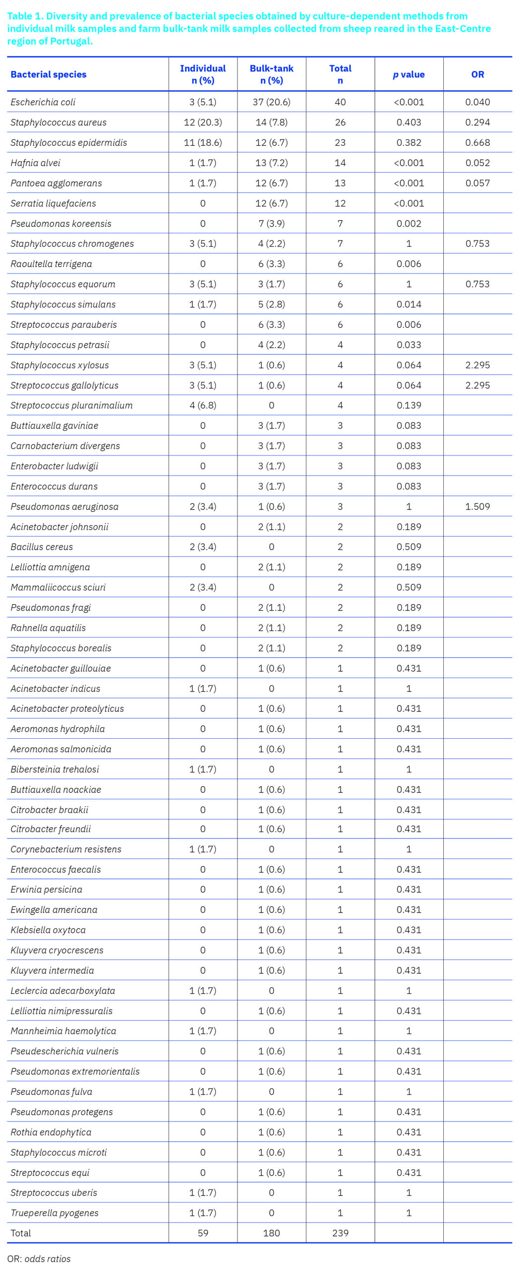
Most of the individual milk samples did not show bacterial growth (59.1%). In the 54 individual milk samples that were culture positive, 22 bacterial species from 14 genera were isolated. In most positive samples, only one bacterial species was isolated (85.2%), but in seven samples (13.0%) and in one sample (1.9%) two and three different bacterial species were obtained, respectively, making a total of 59 isolates recovered from individual milk samples. The most frequently isolated bacteria were S. aureus (20.3%) and S. epidermidis (18.6%). CNS represented 35.6%, followed by S. aureus (20.3%) and Enterobacteriaceae (11.9%) of the bacterial isolates.
On the contrary, only one of 100 (1.0%) of the bulk-tank milk samples did not show bacterial growth. In 40.4% of the samples only one bacterial species was isolated, but in 38.4% and 21.2% of the samples two and three bacterial species were isolated, respectively, making a total of 180 bacterial isolates recovered from the bulk-tank milk samples. The diversity of bacterial species isolated from the bulk-tank was higher than in the individual milk samples, and included 44 species belonging to 23 genera. The most frequently isolated microorganism was E. coli (20.6%). Enterobacteriaceae represented 53.1% of the isolates, followed by CNS (17.9%) and S. aureus (7.8%).
- coli, Hafnia alvei, and Pantoea agglomerans showed ORs well below 1 (0.040; 0.052; 0.057, respectively), indicating a strong association with bulk-tank milk samples and statistically significant differences. Staphylococcus xylosus and Streptococcus gallolyticus had an OR above 1 (2.295), suggesting a higher prevalence in individual milk samples, although these differences were not significant. Most other bacteria had an OR near 1, showing minimal differences between sample type (Table 1).
Susceptibility profile of bacteria isolated from individual and bulk-tank milk samples
AST was performed on 217 bacterial isolates, of which 59 were obtained from individual milk samples and 158 from farm bulk-tank milk samples. The results from the most frequently isolated bacterial species, E. coli, S. epidermidis and S. aureus are presented below.
Escherichia coli
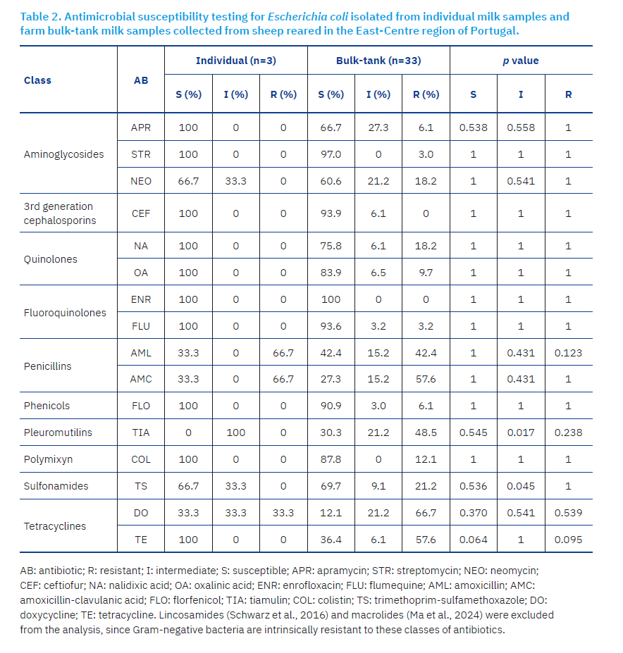
The three E. coli isolates obtained from individual milk samples demonstrated 100% susceptibility to apramycin, streptomycin, ceftiofur, nalidixic acid, oxalinic acid, enrofloxacin, flumequine, florfenicol, colistin, and tetracycline. High rates of resistance (>20%) were observed to three of 16 (18.8%) of the antibiotics tested, namely penicillins (amoxicillin, amoxicillin-clavulanic acid, 66.7%), and tetracyclines (doxycycline, 33.3%). No multi-resistant isolates were obtained from individual milk samples.
The resistance/susceptibility profile of the 33 E. coli isolates obtained from the bulk-tank milk was slightly different from individual milk samples. High rates of susceptibility (>90%) were obtained to 31.3% of the antibiotics tested, namely streptomycin (97.0%), ceftiofur (93.9%), enrofloxacin (100%), flumequine (93.6%), and florfenicol (90.9%). Hight rates of resistance were observed to 37.5% of the antibiotics tested, namely amoxicillin (42.4%), amoxicillin-clavulanic acid (57.6%), tiamulin (48.5%), trimethoprim-sulfamethoxazole (21.2%), doxycycline (66.7%), and tetracycline (57.6%). Further, 21 E. coli isolates (63.6%) from the bulk-tank milk were considered multi-resistant (Table 2).
Statistically significant differences in the profile of susceptibility/resistance of E. coli isolated from individual and bulk-tank milk samples were observed for tiamulin (p=0.017), and trimethoprim-sulfamethoxazole (p=0.045).
Staphylococcus epidermidis
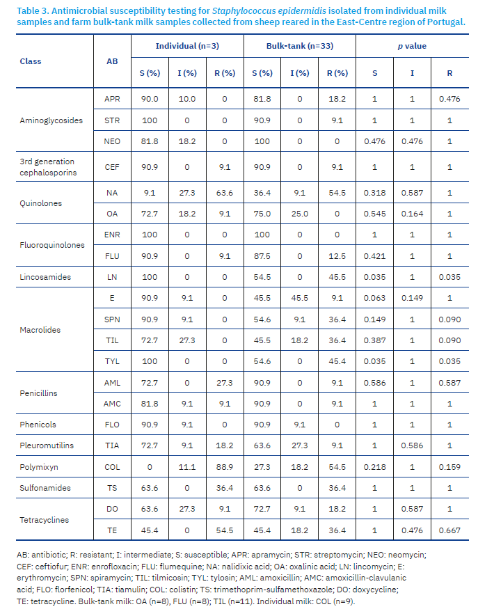
S. epidermidis isolates obtained from individual milk samples demonstrated high rates of susceptibility to 42.9% of the antibiotics tested, namely streptomycin (100.0%), ceftiofur (90.9%), enrofloxacin (100.0%), flumequine (90.9%), lincomycin (100.0%), erythromycin (90.9%), spiramycin (90.9%), tylosin (100.0%), and florfenicol (90.9%). High rates of resistance were observed to 23.8% of the antibiotics tested, namely nalidixic acid (63.6%), amoxicillin (27.3%), colistin (88.9%), trimethoprim-sulfamethoxazole (36.4%), and tetracycline (54.5%). Eight of the 11 (72.7%) S. epidermidis isolates from individual milk samples were multi-resistant.
S. epidermidis isolated from farm bulk-tank milk samples, demonstrated high rates of susceptibility to 33.3% of the antibiotics tested, namely streptomycin (90.9%), neomycin (100.0%), ceftiofur (90.9%), enrofloxacin (100.0%), amoxicillin (90.9%), amoxicillin-clavulanic acid (90.9%), and florfenicol (90.9%). High rates of resistance were observed to 38.1% of the antibiotics tested, namely nalidixic acid (54.5%), lincomycin (45.5%), spiramycin (36.4%), tilmicosin (36.4%), tylosin (45.4%), colistin (54.5%), trimethoprim-sulfamethoxazole (36.4%), and tetracycline (36.4%). Six of the 11 S. epidermidis isolates (54.5%) from the bulk-tank milk samples were multi-resistant (Table 3).
Statistically significant differences were observed in the susceptibility/resistance profile of S. epidermidis isolated from individual and bulk-tank milk samples to lincomycin (p=0.035) and tylosin (p=0.035).
Staphylococcus aureus
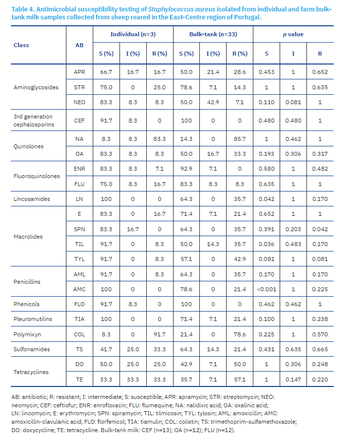
S. aureus isolates obtained from individual milk samples revealed high rates of susceptibility to 38.1% of the antibiotics tested, namely ceftiofur (91.7%), lincomycin (100.0%), tilmicosin (91.7%), tylosin (91.7%), amoxicillin (91.7%), amoxicillin-clavulanic acid (100%), florfenicol (91.7%), and tiamulin (100.0%). Hight rates of resistance were observed to 28.6% of the antibiotics tested, namely streptomycin (25.0%), nalidixic acid (84.6%), colistin (91.7%), trimethoprim-sulfamethoxazole (33.3%), doxycycline (30.8%), and tetracycline (38.5%). Seven of the 12 (58.3%) S. aureus isolates were multi-resistant.
S. aureus isolated from bulk-tank milk samples presented high rates of susceptibility to 14.3% of the antibiotics tested, namely ceftiofur (100%), enrofloxacin (92.3%), and florfenicol (100%). High rates of resistance were observed to 71.4% of the antibiotic tested, namely apramycin (28.6%), nalidixic acid (85.7%), oxalinic acid (35.7%), lincomycin (35.7%), erythromycin (21.4%), spiramycin (35.7%), tilmicosin (35.7%), tylosin (42.9%), amoxicillin (35.7%), amoxicillin-clavulanic acid (21.4%), tiamulin (21.4%), colistin (78.6%), trimethoprim-sulfamethoxazole (21.4%), doxycycline (50.0%), and tetracycline (57.1%). Eleven of the 14 (78.6%) S. aureus isolates obtained from the bulk-tank were multi-resistant.
Statistically significant differences were observed in the rates of susceptibility/resistance of S. aureus isolated from individual and bulk-tank milk samples to the antibiotics lincomycin (p=0.042), spiramycin (p=0.042), tilmicosin (p=0.036), and amoxicillin-clavulanic acid (p<0.001) (Table 4).
Discussion
Milk can be contaminated by bacteria excreted by females with bacterial IMI. However, milk from healthy udders can also be colonised by bacteria transferred from other reservoirs. The direct contact of milk with contaminated surfaces, such as the teat canal, teat skin, and milk equipment may result in milk contamination. On the other hand, feed material, grass, soil, air, litter, drinking and washing water, stable, milker and milk parlour can act as indirect reservoirs of bacteria that can contaminate milk (Rocha et al., 2021). Thus, the microbiological quality of milk reflects the udder health and the subsequent contamination that can occur through direct and indirect reservoirs.
Routine microbiological diagnosis is based on culture-dependent approaches; however, their sensibility is not ideal (Toquet et al., 2021). Indeed, about half of our individual milk samples obtained from ewes with clinical and subclinical mastitis were culture negative. A similar percentage of culture negative samples (50%) was observed in a study that analysed samples of subclinical and clinical mastitis (Makovec and Ruegg, 2003), but low percentages of no growth were reported in samples of clinical (20–44%) (Nevala et al., 2004; Bradley et al., 2007; Koivula et al., 2007; Olde Riekerink et al., 2008) and subclinical mastitis (29–39%) (Bradley et al., 2007; Koivula et al., 2007). The absence of bacterial growth in clinical and, especially, in subclinical mastitis may be attributed to the low concentration of the bacteria in the milk sample, although some studies have observed a lack of growth in samples with a high amount of bacterial DNA (Taponen et al., 2009). The presence of endogenous antibacterial factors in the milk, such as lactoferrin, lysozyme, lactoperoxidase, components of complement, and immunoglobulins (Rainard and Riollet, 2006), working in synergy with milk leukocytes, could decrease the viability of bacteria in culture (Taponen et al., 2009; Barlow, 2011). Inappropriate storage and transport of milk samples, as well as long periods between collection and analysis, can have the same effect. The infection of the mammary gland by bacteria that do not grow in conventional culture media, such as Mycoplasma agalactiae (Hegde et al., 2014; Rahimabadi et al., 2017) and the infection by virus, such Maedi-Visna virus, which is highly prevalent in Portugal (Kalogianni et al., 2020; Jacob-Ferreira et al., 2023a, 2023b) could explain the absence of bacterial growth in sheep milk with suspected IMI. Several authors have suggested that the rapid clearance of some bacteria from the mammary gland, as is the case of E. coli, could explain some negative culture results (Olde Riekerink et al., 2008). Experimental models have shown that in bovine mastitis, clinical signs of inflammation, namely the SCC, remain high for days after infection clearance. Finally, mammary inflammation may be non-infectious, and instead may be caused by chemical, physical or traumatic causes (Taponen et al., 2009).
The community of bacteria that can be found in milk is very diverse and farm-specific (Castro et al., 2019), comprising species that assist dairy fermentation and/or promote consumer health, and pathogenic species that can cause IMI and human diseases (Quigley et al., 2013). Furthermore, culture-independent methods allowed the identification of a community of resident bacteria within the teat duct that could provide some protection against invading microorganisms (commensal flora), but that under certain circumstances can reach the mammary parenchyma, acting as opportunistic (Castro et al., 2019; Katsafadou et al., 2019; Toquet et al., 2021). Several studies suggest that mastitis is the result of an imbalance of the mammary microbiota (mammary dysbiosis) rather than the action of a unique pathogen (Contreras and Rodríguez, 2011; Castro et al., 2019; Toquet et al., 2021). In this context, the distinction between commensal, pathogenic for sheep and humans, and technological important bacteria is not always clear.
This study employed culture-based methods to isolate bacterial species from ewe milk samples, which were then identified to the species level using MALDI-TOF technology. Bacteria isolated from milk samples are traditionally identified to the species level by means of biochemical tests or commercial biochemical galleries. However, these methods often fail in correctly identifying bacterial species of veterinary interest, because they have been optimised for human bacterial strains (Rosa et al., 2022). MALDI-TOF has been successfully applied in cow (Nonnemann et al., 2019; Conesa et al., 2020) and small ruminant milk (Vasileiou et al., 2019; Knuth et al., 2022; Rosa et al., 2022), allowing for accurate and rapid bacterial identification, which is very advantageous when the purpose is the prompt treatment or disposal of affected animals. In the present study, a total of 55 bacterial species belonging to 31 genera were identified by MALDI-TOF. In addition to S. aureus, eight species of CNS, four species of Streptococcus, four species of Pseudomonas, 16 species of Enterobacteriaceae, among others were identified by MALDI-TOF with high log scores, demonstrating the power of this technology in identifying bacterial species of veterinary interest.
According to previous studies, S. aureus is the primary etiological agent of clinical mastitis in ewes, having been isolated from up to 65.3% of clinical mastitis cases (Bergonier et al., 2003; Mørk et al., 2007; Arsenault et al., 2008; Vasileiou et al., 2019), whereas CNS species, namely S. chromogenes, S. epidermidis, S. simulans and S. xylosus are considered the primary etiological agents of subclinical mastitis, comprising up 70% of the isolates (Gelasakis et al., 2015; Vasileiou et al., 2019). In the present study, where milk samples were collected from ewes with clinical and subclinical mastitis, the most frequently isolated microorganisms were S. aureus (20.3%) and CNS (35.6%), although 11.9% of the bacterial isolates belonged to the Enterobacteriaceae family. The main sources of Staphylococcus spp. are the hands of milkers (Albenzio et al., 2003; Vasileiou et al., 2018), nasopharynx of suckling lambs (Albenzio et al., 2003; Gougoulis et al., 2008), and the teat duct and udder skin (Fragkou et al., 2007; Mavrogianni et al., 2007), which reach the mammary gland during milking or suckling, whereas Enterobacteriaceae are environmental pathogens that invade the mammary gland between milkings (Bradley et al., 2007; Vanderhaeghen et al., 2014).
Considering the European legislation (EP, 2004) the mesophilic bacterial count (MBC) is used to assess the bacteriological quality of raw milk. However, the EU legislation does not require mandatory investigation of pathogenic bacteria present in raw milk. Therefore, raw milk and artisanal cheeses produced with raw milk may contain pathogenic bacteria (Almeida et al., 2007; Výrostková et al., 2021; Praça et al., 2023), representing a potential risk, particularly for vulnerable groups of consumers (EFSA and ECDC, 2021; Nüesch-Inderbinen et al., 2021). Thus, auto-control programmes aimed to evaluate the microbiological quality of bulk-tank milk are essential (Lianou et al., 2021; Parco et al., 2021). In the present study, 99% of the bulk-tank samples presented bacterial grow. The diversity of bacterial species isolated from the bulk-tank milk was higher than in individual milk samples, with Enterobacteriaceae prevailing. Furthermore, a significantly higher prevalence of E. coli, Hafnia alvei, and Pantoea agglomerans was observed in bulk-tank milk samples than in individual milk samples. These results indicate that contamination of bulk-tank milk is not only of ewe origin, rather than from other sources, namely from human and environmental origins (Albenzio et al., 2003; Lianou et al., 2021). In agreement with our results, a study performed on four sheep farms in Eastern Hungary observed that bulk-tank milk samples contained up to 10,000 times as many bacteria as did their individual raw milk counterparts (Pulina et al., 2018). According to the authors, the elevation in bacterial concentration in bulk-tank milk reflects the level of hygiene during milking and milk handling, including storage. Previous studies had already observed that bacterial count increases significantly in raw milk during storage at 4°C (de Garnica et al., 2011).
Some of the bacteria overrepresented in the bulk-tank milk were identified as part of the microbiome of raw milk cheese, namely some Enterobacteriaceae (Salamandane et al., 2024). Despite the great diversity of bacteria present in bulk-tank milk, cheese made of raw milk is considered a microbiologically safe product, with no records of food-borne outbreaks related to the consumption of contaminated Portuguese cheeses. The reduction of pH during cheese manufacturing due to the metabolic activity of lactic acid bacteria, nutrient depletion, release of antimicrobial compounds, water activity decrease and the low temperature at ripening stages promote a hostile environment for bacteria development, promoting cheese safety (Rampanti et al., 2022; Rocha et al., 2023).
Understanding the antimicrobial susceptibility profile of bacteria involved in IMI is crucial for effective treatment and management of the infection (Iancu et al., 2024). Furthermore, the AST results can be used to generate epidemiological data, allowing for the development of antibiotic prescription protocols based on local/regional antimicrobial sensitivity trends (EU, 2015), and to design public policies to combat the development and spread of AMR. In this study, we provided data on AST of 217 bacterial isolates, obtained from individual and bulk-tank milk samples.
E. coli is intrinsically susceptible to almost all clinically relevant antibiotics, except to lincosamides (Schwarz et al., 2016) and macrolides (Ma et al., 2024), but have a great capacity to accumulate resistance genes, mostly through horizontal gene transfer (Poirel et al., 2018). E. coli isolates obtained from individual and bulk-tank milk samples were resistant to penicillins and tetracyclines. E. coli strains resistant to penicillins through the production of extended-spectrum b-lactamases are clinically relevant in veterinary medicine and were previously described in ewe milk samples (Solomakos et al., 2009; Poirel et al., 2018; Obaidat et al., 2023; Kürekci et al., 2024). Several studies described strains of E. coli resistant to tetracyclines (Solomakos et al., 2009; Obaidat et al., 2023; Kürekci et al., 2024). Both penicillins and tetracyclines are widely used in veterinary medicine, imposing a selective pressure that has culminated in the spread of E. coli resistant strains (Poirel et al., 2018; Kürekci et al., 2024). E. coli isolated from bulk-tank milk revealed a wide range of resistance, including penicillins and tetracyclines, but also pleuromutilins and sulfonamides.
A major characteristic of most Staphylococcus spp., including S. aureus, is their extended ability to rapidly develop resistance to antimicrobial agents (Vasileiou et al., 2019). Although IMI caused by CNS is generally less serious than those caused by S. aureus, CNS has a negative impact on the quality and quantity of milk and SCC, producing persistent subclinical infections. On the other hand, CNS are considered reservoirs of antimicrobial resistant genes (ARG) for S. aureus, which are generally more virulent and have greater clinical importance for animals and humans (Barrero-Domínguez et al., 2019; de Souza Santo et al., 2020). In this study, S. epidermidis and S. aureus isolated from individual milk samples revealed high rates of resistance to 23.3% and 28.6% of the antibiotics tested, whereas the same bacteria isolated from bulk-tank milk samples revealed high rates of resistance to 38.1% and 71.4% of the antibiotics. More than 50% of the S. epidermidis and S. aureus isolates were multi-resistant. However, the Staphylococcus spp. resistance profile does not differ much from that observed in other studies (Parco et al., 2021; Iancu et al., 2024), although it is difficult to establish comparisons due to the different antibiotics tested and laboratory methodologies used.
Together, these results indicate that the resistance profile of the main bacteria isolated from sheep milk, namely E. coli and Staphylococcus spp., seems to be more worrying in bulk-tank milk than in milk from sheep with clinical and subclinical mastitis. Although the individual and bulk-tank milk samples analysed in this study are from different farms and therefore do not allow direct extrapolation, these results seem to indicate that bulk-tank milk “accumulates” resistance. Since the bulk-tank stores the milk of all lactating ewes in the farm, it is expected to act as a reservoir of antimicrobial resistant bacteria (ARB) and ARG from animals, but also from external sources, which can be transferred vertically during bacteria division and horizontally between bacteria through mobile genetic elements (Schwarz et al., 2017). Transference of ARB and ARG to humans through the food chain is of great concern. Recent studies suggest the presence of methicillin resistant Staphylococcus (MRSA) in unpasteurized cow and sheep milk and dairy products, including cheeses (Freitas Ribeiro et al., 2020; Regecová et al., 2021; Výrostková et al., 2021; Salamandane et al., 2024), which constitute another route of AMR dissemination to humans (Vasileiou et al., 2019).
Despite the relatively low number of isolates of some bacterial species studied, this study investigated for the first time the resistance/susceptibility profile of bacteria obtained by culture-dependent approaches in milk of ewe with suspected IMI and bulk-tank milk produced in the East-Centre region of Portugal. Due to the scarcity of epidemiological data available, the results obtained are essential for guiding appropriate antibiotic therapy, monitoring resistance patterns, and developing strategies to mitigate the spread of AMR in this region.
To improve the hygienic quality of milk and reduce the emergence of antimicrobial resistance (AMR), it is essential to implement measures that allow for a reduction of the use of antibiotics in food-producing animals, through structural improvements on farms, particularly in milking parlours, raising awareness to strengthen biosecurity measures and medical prophylaxis, and controlling access to veterinary medicines.
Conclusions
Most of the individual milk samples from ewes with suspected IMI did not show bacterial growth, while virtually all bulk-tank milk samples were culture positive.
CNS, S. aureus and Enterobacteriaceae represented 35.6%, 20.3% and 11.9% of the bacterial isolates obtained from sheep individual milk samples, respectively, while in bulk-tank milk Enterobacteriaceae represented the most frequently isolated microorganisms (53.1%), followed by CNS (17.9%) and S. aureus (7.8%).
Higher rates of resistance were obtained for E. coli, S. epidermidis and S. aureus isolates obtained from the bulk-tank than for bacterial strains obtained from individual milk samples.
E. coli from individual milk samples revealed high rates of resistance to penicillins and tetracyclines, whereas S. aureus revealed high rates of resistance to aminoglycosides, polymyxin, sulfonamides and tetracycline, maintaining high levels of susceptibility to third generation cephalosporins, lincosamides, some macrolides, penicillins, phenicols and pleuromutilins.
Author Contributions: Conceptualization, M.A.P.; methodology, M.A.P and T.F.; validation, M.A.P, T.F and A.L.B.; formal analysis, M.A.P, A.L.B and M.M.; investigation, A.L.B, R.C., F.E., J.M., C.G. and C.S.; resources, M.A.P; data curation, J.M.; writing—original draft preparation, M.A.P and A.L.B.; writing—review and editing, R.C, F.E., J.M., C.N., A.C.M., C.G., C.C., C.S., M.M. and H.V.; supervision, M.A.P.; project administration, M.A.P; funding acquisition, M.A.P. All authors have read and agreed to the published version of the manuscript.
Funding: This work was funded by the RumiRes project – “Vigilância epidemiológica de resistências antimicrobianas e resíduos medicamentosos em Pequenos ruminantes da Região Centro” (Ref. PRR-C05-i03-I-000190); Science and Technology Foundation through funds for GHTM-UID/04413/2020 e LA-REAL—LA/P/0117/2020 and CERNAS UIDB/00681/2020.
Institutional Review Board Statement: Ethical review and approval of this study were waived because all biological samples were collected for diagnostic purposes by farm veterinarians.
Acknowledgments: The authors acknowledge the producers, cheese factories and private veterinarians and veterinary nurses (Luís Lourenço, Pedro Carreira, Daniel Correia, Susana Mendes, Diogo Themudo, João Castelo Branco, Pedro Caseiro), who contributed to this work by collecting milk samples for analysis.
References [… show]
Raznolikost i profil antimikrobne rezistencije bakterija izoliranih kod ovaca s mastitisom i iz mlijeka iz spremnika u istočno-središnjem Portugalu
Maria AIRES. PEREIRA1,2,3* (corresponding author), mapereira@esav.ipv.pt, orcid.org/0000-0002-4054-9124; Alexandra LAMEIRA BAPTISTA1,2,4, alexabaptista@esav.ipv.pt, orcid.org/0009-0002-8137-1139; Mariana ROSÁRIO1, mariana.rosario04@gmail.com, orcid.org/0009-0001-6429-3573; Rita Marisa da SILVA CRUZ 1,5,6, rcpaiva@esav.ipv.pt, orcid.org/0000-0002-5767-7835; Fernando Alexandre de ALMEIDA ESTEVES1,2 festeves@esav.ipv.pt, orcid.org/0000-0003-0589-0746; João MADANELO1; joao.madanelo@gmail.com; Carmen Lucia de VASCONCELOS NOBREGA1,7 cnobrega@esav.ipv.pt, orcid.org/0000-0003-3941-799X; Ana Cristina PAIS MEGA DE ANDRADE 1,2, amega@esav.ipv.pt, orcid.org/0000-0002-9913-875X; Carla GARCIA1, cgarcia@esav.ipv.pt; Catarina COELHO1,2, ccoelho@esav.ipv.pt, orcid/org0000-0002-5272-7303; Carla Sofia AREDE DOS SANTOS 1, casarede@esav.ipv.pt, orcid.org/0000-0001-5908-8500; Madalena MALVA8 malva@esav.ipv.pt, orcid.org/0000-0003-4982-0184; Helena VALA1,2,9 h.vala@sc.ipv.pt, orcid.org/0000-0001-6829-4867; Tiago FERREIRA10 tiagoandreferreir@gmail.com.
1Instituto Politécnico de Viseu, Escola Superior Agrária de Viseu, Av. Dr. António Almeida Henriques, 3500-631 Viseu, Portugal;
2CERNAS-IPV Research Centre, Instituto Politécnico de Viseu, Campus Politécnico, Repeses, 3504-510 Viseu, Portugal;
3Global Health and Tropical Medicine, GHTM, Associate Laboratory in Translation and Innovation Towards Global Health, LA-REAL, Instituto de Higiene e Medicina Tropical, IHMT, Universidade NOVA de Lisboa, UNL, Rua da Junqueira 100, 1349-008 Lisboa, Portugal;
4 Universidade de Trás-os-Montes e Alto Douro, Quinta de Prados 5000-801, Vila Real, Portugal;
5Epidemiology Research Unit (EPIUnit), Instituto de Saúde Pública da Universidade do Porto, Rua das Taipas 135, 4050-600, Porto, Portugal;
6Laboratory for Integrative and Translational Research in Population Health (ITR), Rua das Taipas 135, 4050-600, Porto, Portugal;
7Center for the Research and Technology of Agro-Environmental and Biological Sciences (CITAB), University of Trás-os-Montes e Alto Douro, Quinta de Prados, Edifício Reitoria, 5000-801, Vila Real, Portugal;
8Instituto Politécnico de Viseu, Escola Superior de Tecnologia e Gestão de Viseu, Avenida Cidade Politécnica, 3504-510, Viseu, Portugal;
9Veterinary and Animal Research Centre (CECAV), UTAD, Associate Laboratory for Animal and Veterinary Sciences (AL4AnimalS), Quinta de Prados, Apartado 1013, 5000-801, Vila Real, Portugal;
10Australian Laboratory Services (ALS), Zona Industrial de Tondela Lote 6, 3460-070, Tondela, Portugal.Mikrobiološka kvaliteta mlijeka odražava zdravlje vimena i naknadnu kontaminaciju koja može nastati putem izravnih i neizravnih rezervoara. Cilj ovog istraživanja bio je identificirati bakterijsku raznolikost u uzorcima mlijeka ovaca s mastitisom i u mlijeku iz spremnika te istražiti njihove profile osjetljivosti i otpornosti na antibiotike. Analizirano je ukupno 232 uzorka mlijeka. Iz pojedinačnih uzoraka mlijeka izolirane su 22 bakterijske vrste, među kojima se ističu Staphylococcus aureus (20,3%) i Staphylococcus epidermidis (18,6%). Raznolikost bakterijskih vrsta izoliranih iz spremnika bila je veća i obuhvaćala je 44 vrste, s prevladavanjem E. coli (20,6%), S. aureus (7,8%) i Hafnia alvei (7,2%). Testiranje osjetljivosti na antimikrobne lijekove provedeno je na 217 bakterijskih izolata. E. coli izdvojena iz pojedinačnih uzoraka mlijeka pokazala je visoke stope otpornosti na peniciline i tetracikline, dok je S. aureus pokazao visoke stope otpornosti na aminoglikozide, polimiksin, sulfonamide i tetraciklin, ali i visoku razinu osjetljivosti na cefalosporine treće generacije, linkozamide, neke makrolide, peniciline, fenikole i pleuromutiline. Profil otpornosti bakterija izoliranih iz mlijeka iz spremnika zabrinjavajući je u odnosu na uzorke mlijeka ovaca s mastitisom. Ovi rezultati ukazuju na potrebu za strukturnim poboljšanjima na farmama i podizanjem svijesti o jačanju biosigurnosti, medicinskoj profilaksi i kontroli pristupa veterinarskim lijekovima kako bi se poboljšala kvaliteta mlijeka i smanjila upotreba antibiotika.
Ključne riječi: Escherichia coli; koagulaza-negativni Staphylococcus; Staphylococcus aureus; intramarne infekcije; laktofriz; određivanje antimikrobne osjetljivosti.