
Hock Arthritis in Pet Chickens (Gallus gallus domesticus): Physical, Cytological and Bacteriological Study of Synovial Fluid
S. Derbal
Said DERBAL1 derbal.said@crsp.dz, orcid.org/0000-0002-1805-8775.
1Pharmaceutical Sciences Research Center, 25016 Ali Mendjeli, Constantine, Algeria
![]()
https://doi.org/10.46419/cvj.57.5.2
Abstract
Hock arthritis is a pathology with significant repercussions on the health of pet chickens, and on the economics of the poultry sector. The objective of this study was to detail the physical, cytological, and bacteriological characteristics of synovial fluid from hocks of 52 pet chickens affected by arthritis. The appearance of synovial fluid was assessed in terms of volume, colour, turbidity and viscosity. The mucin clot test was performed. Synovial fluid smears were stained with May-Grunwald-Giemsa to perform differential cell counts. Isolated bacteria were identified by standard microbiological and biochemical tests. Analysis of the synovial fluid samples revealed variations in their physical characteristics. The majority of samples exhibited a volume of 200 µL, slight turbidity, a bloody colour and decreased viscosity. The number of erythrocytes, platelets and white blood cells varied depending on the sample for cytological parameters. One sample showed an absence of leukocytes. Synovial fluids infected with Staphylococci showed a higher number of leukocytes per field compared to those infected with Enterobacteriaceae. Bacteriological analysis revealed that 65.38% of synovial fluid samples were negative for bacteria, while 34.61% were positive, of which Staphylococcus aureus (S. aureus) (35%), Staphylococcus epidermidis (S. epidermidis) (40%), Escherichia coli (E. coli) (5%) and Salmonella enterica (S. enterica) (20%). Synovial fluid in hock arthritis shows variable physical and cytological characteristics, reflecting changes in cell types and fluid properties. In pet chickens, the main bacterial agents identified are S. aureus, S. epidermidis, E. coli and S. enterica. These findings provide essential information for veterinarians and poultry practitioners, aiding in joint health assessment, early diagnosis and timely treatment.
Key words: hock arthritis; pet chickens; synovial fluid; cytology; bacteriology.
Introduction
Arthritis is a health issue in pet chickens, especially when it leads to lameness, resulting in significant welfare problems. Several pathogens, such as bacteria, mycoplasmas and viruses, can be responsible for this condition. Infectious arthritis can result from the direct introduction of microorganisms within the intra-articular space, spread from periarticular infection or hematogenous spread (Francoz et al., 2005). The severity of joint destruction depends on the bacterial species and the duration of infection, with each type of arthritis-inducing bacteria according to its virulence factors (Soontornvipart et al., 2003). Joint diseases in chickens are typically suspected based on clinical signs such as lameness, reluctance to walk, joint swelling or heat, pain on palpation, reduced feed intake and decreased weight gain. While physical examination can detect structural changes, they often cannot distinguish infectious from non-infectious causes. Synovial fluid analysis provides definitive confirmation of joint infection, allowing evaluation of physical characteristics and cytological parameters. Additionally, synovial fluid can be cultured to identify causative microorganisms, differentiating infectious or non-infectious inflammatory conditions and guiding targeted treatment (McIlwraith et al., 2001).
Synovial fluid is produced by the synovial membrane and synoviocytes and contains hyaluronic acid and proteins that give it its viscosity. Appearing as a highly viscous plasma dialysate, synovial fluid provides lubrication, nutrition, load-bearing and shock absorption within the joints (Nazifi et al., 1998). Healthy synovial fluid is clear, pale yellow, viscous, and does not form clots (AbdEllah et al., 2012). Disturbances or alterations in the exchange of substances between the vascular and lymphatic systems and synovial fluid can result from various diseases (Nazifi et al., 1998). Synovial fluid analysis is essential to identify cytological and physical abnormalities in inflammatory conditions, whether suppurative or not, as well as in cases of haemorrhages, malignant tumours or infectious diseases (Moraes et al., 2010). It remains the standard diagnostic method for degenerative, inflammatory, mechanical, traumatic, metabolic and tumoral joint disorders (Al-Rukibat et al., 2006), especially in lame animals with joint effusion (AbdEllah et al., 2012). According to Boon (1997), synovial fluid assessment is performed through physical and cytological analyses.
Early diagnosis and treatment of arthritis in pet chickens are of paramount importance, and synovial fluid analysis is essential to obtain crucial information on the aetiology, inflammatory status, appropriate treatment and progression of the condition. Notably, synovial fluid cytology, considered an essential ancillary test, helps differentiate infectious from non-infectious arthritis (Rohde et al., 2000). Similarly, the differential leukocyte count helps to identify the causes of arthritis by determining the predominant type of white blood cell (AbdEllah et al., 2012).
Although numerous studies have explored synovial fluid analysis in mammals (Nazifi et al., 1998, 2005; Ismail et al., 2007; Waseem et al., 2016), the literature addressing synovial fluid pathology in chickens and the values associated with its analysis currently is both outdated and limited. Most existing research focuses on the synovial fluid of the hock joint because it is easy to access, yields sufficient fluid for routine sampling (Corr et al., 2003), and can be reached with minimal trauma (Morrow et al., 1997). Data regarding hock arthritis in pet chickens are lacking. Analysis of synovial fluid in pet chickens offers a valuable diagnostic tool, as it allows for the rapid detection of joint inflammation, the identification of bacterial or viral infections, and the assessment of the severity of osteoarticular lesions. Therefore, the objective of our study was to describe the results of physical, cytological and bacteriological analysis of joint synovial fluid from pet chickens with hock arthritis.
Materials and methods
Chickens with arthritis
Synovial fluid samples were collected from 52 pet chickens with hock arthritis, aged 50 to 60 days and belonging to different farms in the Sétif Province (Algeria). These chickens are fed a commercial, locally sourced diet. Before selection, live chickens underwent a standing and locomotion assessment. A physical examination, including joint palpation, was performed. All chickens exhibited lameness and swelling of the hock joints and were included in our study.
Synovial fluid collection
Prior to collection, each leg was disinfected with a povidone-iodine solution (10%) (Betadine®) and surgical alcohol at the hock joint for better disinfection. To aspirate the synovial fluid, a sterile 18-gauge, 1.5-inch needle attached to a sterile 3 mL syringe was inserted through the caudolateral aspect of the hock joint into the joint space. Synovial fluid was carefully collected to avoid increasing stress and pain in the chickens, and then major quantities of synovial fluid were placed in tubes containing calcium ethylenediamine tetra acetic acid (EDTA) for physical and cytological analyses (Waseem et al., 2016). A few drops of synovial fluid were poured into sterile Eppendorf tubes for bacterial culture. If only a small amount of fluid was obtained, a few air-dried smears were made on slides.
Synovial fluid analysis
Physical analysis
Physical analysis of the synovial fluid was performed by determining the volume of aspirated fluid, its colour, turbidity, viscosity and the presence of old blood or fresh clots, immediately after collection (Corr et al., 2003; Ismail et al., 2007; Moraes et al., 2010). The small amount of synovial fluid collected from pet chickens did not allow for the analysis of other parameters.
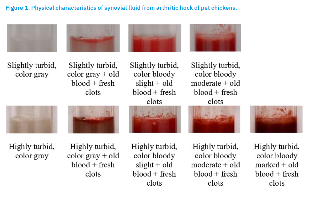
The colour of healthy synovial fluid is pale yellow, and colour variations in pathological synovial fluid has potential diagnostic significance. Grey fluid may indicate chronic inflammation or degenerative joint disease; slightly bloody indicates minor haemorrhage possibly from mild trauma, joint aspiration or early inflammatory changes; moderately bloody suggests more pronounced haemorrhage and could indicate severe trauma, vascular damage or moderate septic arthritis; markedly bloody is strongly suggestive of significant trauma or advanced septic arthritis; old blood indicates prior haemorrhage and could suggest a chronic or previously resolved joint injury or inflammation; while fresh clots indicate active or recent bleeding; often associated with acute trauma, acute septic arthritis or vascular rupture.
The turbidity of healthy synovial fluid is clear with no visible particles, while the turbidity of pathological synovial fluids is classified as: 1) slightly turbid: slight cloudiness, particles barely visible; 2) moderately turbid: noticeable cloudiness, with some suspended material; or 3) highly turbid: opaque, with dense particulate matter.
Fluid viscosity was assessed subjectively using the string test, in which the fluid was stretched between a finger and thumb. Viscosity was considered decreased if it extended less than 2 cm or dropped like water, normal if it extended 2 cm, and increased if it extended more than 2 cm (Corr et al., 2003).
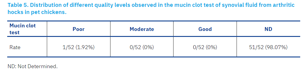
The mucin clot test was performed for fluids of sufficient volume (≥ 500 µL) by adding 0.1 mL 7N glacial acetic acid to 4 mL distilled water in a test tube. The resulting solution (2.5%) was gently added to a 0.5 mL aliquot of synovial fluid, ensuring that the sample did not come into contact with the wall of the test tube, in order to ensure a homogeneous and unbiased chemical reaction, necessary for correct interpretation of the test (Moraes et al., 2010). Interpretation of the results followed specific criteria: poor (friable clot, easily broken up by shaking, surrounded by cloudy fluid), moderate (less compact mass with flaps in cloudy solution) and good (a firm, tight mass in clear solution) (Moraes et al., 2010).
Cytological analysis
Synovial smears were prepared on slides, air-dried and then stained with May-Grunwald-Giemsa (Sigma-Aldrich®) for cytological analysis. The stained slides were examined under low magnification (×10) to identify areas of increased cellularity or areas with different staining characteristics which indicates the presence of different types of cells. After selecting a well-stained area, cell counting began progressively from one side to the other. Differential cell counts were performed manually on the synovial smears by examining 50 high-power fields (×1000) per slide (the number of leukocytes/high-power field; leukocytes/HPF) (Corr et al., 2003). This high magnification (×1000) facilitated the identification and relative counting of different cell types (Corr et al., 2003). This cytological analysis was performed after photomicroscopy of the synovial smears using a Euromex® optical microscope equipped with a camera and Image Focus Plus V2 software.
Microbiological Analysis
The collected synovial fluids were enriched in brain heart infusion (BHI) medium (BIOKAR®, France) at a 1:10 dilution and incubated at 37 ± 0.5°C for 24 ± 1 h in an incubator under aerobic conditions. After enrichment, 100 µL broth culture was plated onto Chapman agar (BIOKAR®, France), MacConkey agar (BIOKAR®, France), and blood agar (BIOKAR®, France) plates using a sterile loop and cultured aerobically at 37 ± 0.5 °C for 24–48 h. Synovial fluids were also inoculated into 2 mL mycoplasma broth (BIOKAR®, France) for the isolation of mycoplasmas, with incubation at 37 ± 0.5 °C under 5% CO2 for 3 days.
Bacteria were further identified using standard microbiological tests, including Gram staining, catalase and oxidase tests, motility assessment, and haemolysis pattern. Biochemical identification was performed using API Staph and API 20E strips (bioMérieux®, France), with results interpreted using a technical data sheet according to the manufacturer’s instructions. Mycoplasmas were confirmed by observing colony morphology (typical fried-egg morphology) on selective agar (mycoplasma-specific agar) and, when necessary, by biochemical tests such as glucose fermentation and arginine hydrolysis.
Results
Physical analysis
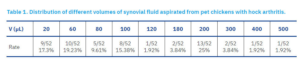
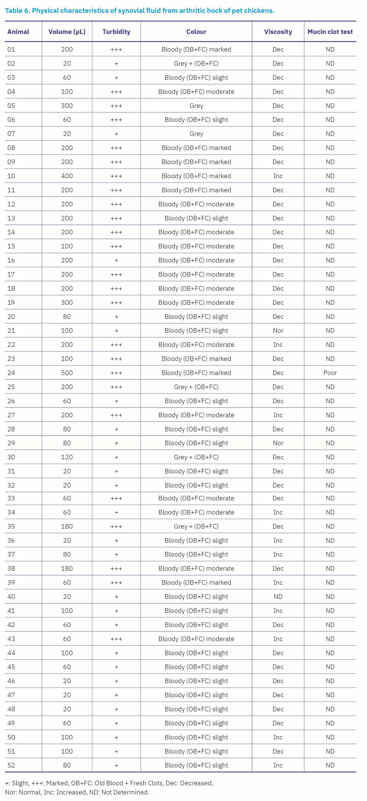
Regarding the volume of synovial fluid samples, the majority (13/52; 25%) had a volume of 200 µL, followed by 60 µL (10/52; 19.23%), 20 µL (9/52; 17.3%), 100 µL (8/52; 15.38%), 80 µL (5/52; 9.61%), 180 µL (2/52; 3.84%), 300 µL (2/52; 3.84%), 120 µL (1/52; 1.92%), 400 µL (1/52; 1.92%) and 500 µL (1/52; 1.92%) (Table 1).
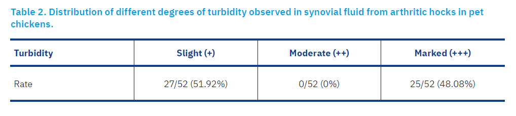
The proportion of synovial fluid samples with slight turbidity (27/52; 51.92%) was slightly higher than that of samples with marked turbidity (25/52; 48.08%). None of the examined samples exhibited a moderate degree of turbidity, as shown in Table 2.
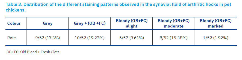
Only two staining patterns were observed in the synovial fluid samples, the majority of which (46/52; 88.46%) exhibited a bloody colour varying in intensity (slight: 24/52; 46.15%, moderate: 14/52; 26.92% and marked: 8/52; 15.38%) with the concomitant presence of old blood and fresh clots. Only 6 of 52 samples (11.53%) exhibited a grey colour, of which 4 (7.69%) contained old blood and fresh clots, as shown in Table 3.
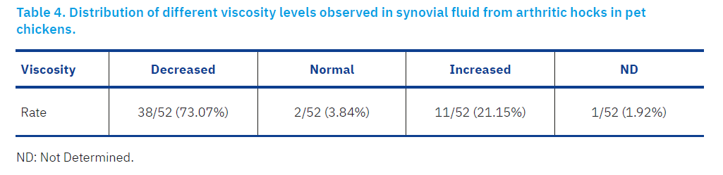
Synovial fluid viscosity was decreased in the majority of samples (38/52; 73.07%), while it was increased in 11/52 samples (21.15%). 2/52 samples (3.84%) presented a viscosity considered normal, as shown in Table 4.
The mucin clot test was performed on a single sample of adequate volume (500 µL), and the results were rated as poor, as shown in Table 5. Table 6 shows the physical characteristics of synovial fluid from arthritic hock of pet chickens. Figure 1 illustrates the results of the physical analysis of synovial fluid conducted in this study.
Cytological and bacteriological analysis
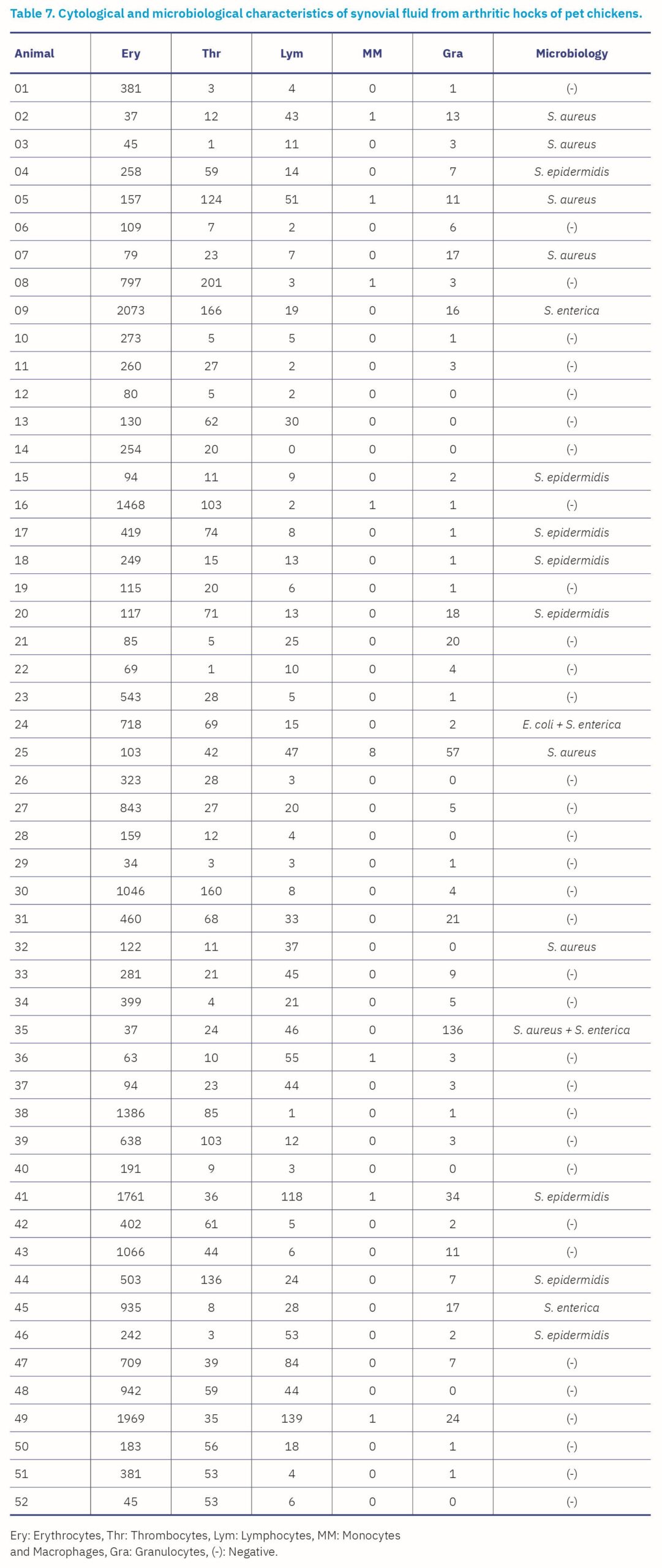
All synovial fluid samples revealed a substantial number of erythrocytes, ranging from 34 to 2073, while the platelet count ranged from 1 to 201. One synovial fluid sample showed a complete absence of lymphocytes, although another had a count as high as 139. Monocytes and macrophages were absent in 44 samples, while one sample had 8 of these cells present. Granulocytes (heterophiles, eosinophils and basophiles) were absent in 9 samples, while one sample contained 136 of these cells (Table 7).
The case of arthritis with coinfection by S. aureus and S. enterica recorded the highest count of leukocytes/HPF, with a value of 182 leukocytes/HPF. Concurrently, one sample showed the absence of bacterial infection and leukocytes. Synovial fluids infected with staphylococci (S. aureus and S. epidermidis) had a count of leukocytes/HPF ranging from 9 to 112, while those infected with enterobacteria had a count ranging from 35 to 45 leukocytes/HPF, as shown in Table 7.
Regarding bacteriological analyses, all samples were negative for mycoplasma. For other bacterial species, 34 of 52 samples (65.38%) were negative, while 18 samples (34.61%) were positive. Among these, 20 bacterial isolates were identified, including S. aureus (7/20; 35%), S. epidermidis (8/20; 40%), E. coli (1/20; 5%) and S. enterica (4/20; 20%), as shown in Table 8.

Discussion
Hock arthritis is a prevalent disease in chicken farms worldwide, causing lameness, inappetence, weight loss and even mortality, resulting in significant economic losses linked to decreased zootechnical performance, increased veterinary costs and degradation of animal welfare. Studies specifically aimed at characterising the physical and cytological properties of synovial fluid from arthritic hocks in pet chickens, as well as identifying the bacterial agents involved, are largely lacking. Therefore, this study focused on exploring the physical, cytological and bacteriological characteristics of synovial fluid in this joint disease in pet chickens.
All synovial fluid samples exhibited pathological changes, illustrated by significant variations in physical, cytological and bacteriological characteristics. Cytological analysis revealed an increased number of leukocytes, including lymphocytes and granulocytes, in all samples positive in bacteriological analysis except sample 32, as well as in some bacteriologically negative samples. The increased presence of leukocytes indicates the exudative properties of the synovial fluid of infected arthritic joints, while the coexistence of erythrocytes with a high number of leukocytes suggests haemorrhages related to infectious vascular lesions in inflamed joints. The abundant microorganisms in synovial fluid can lead to the destruction of the synovial membrane and articular cartilage (Van Pelt, 1974). It is also suggested that haemorrhages in samples with a high number of erythrocytes with a very low number of leukocytes may result from physical trauma. Massive degenerative and inflammatory reactions of the synovial membrane usually lead to the leakage of microorganisms from adjacent infected tissues or from the systemic circulation into the synovial fluid. This causes simultaneous alterations in physical and cytological values, which has diagnostic importance (Paul et al., 2012).
The physical and cytological analyses in this study were consistent with those of Paul et al. (2012), who reported that arthritic synovial fluid in chickens showed a yellowish and cloudy appearance, with variable amounts of flocculent material and clot formation, poor mucin precipitate quality, friable masses in a cloudy, yellow solution, and elevated erythrocyte and leukocyte counts with increased heterophils and monocytes.
It is important to note that physical and cytological parameters of synovial fluid vary among animal species. In birds, they are influenced by breed, age, and physiological status (AbdEllah et al., 2012). These physical and cytological variations in synovial fluid have also been documented in several studies conducted in ruminants with arthritis. Nazaal et al. (2005) performed cytological analyses of synovial fluid in sheep (n = 115) and goats (n = 45), showing variations in the mucin coagulation test, an increase in the total number of leukocytes and the percentage of neutrophils. In another study of 20 synovial fluid samples from clinical cases of arthritis in goats, the appearances were varied, with a significant increase in volume and a significant decrease in viscosity, and variable turbidity, with a predominance of neutrophils in 40% of cases and lymphocytes and monocytes in 60% of cases (Waseem et al., 2016). Cytological analysis of 28 dromedary camels with severe joint disorders revealed a marked increase in both white blood cell and erythrocyte counts, with white cells predominating in the synovial fluid (Ismail et al., 2007). Furthermore, analysis of normal synovial fluid from 43 adult camels revealed a pale, creamy, and clear appearance, with no debris or clot formation observed at room temperature, while the mucin clot test yielded normal results in all samples (Nazifi et al., 1998).
The identification of S. aureus (7/20; 35%), S. epidermidis (8/20; 40%), E. coli (1/20; 5%), and S. enterica (4/20; 20%) in pet chicken arthritis cases in this study highlights the need for special attention from practicing veterinarians. This vigilance is crucial to take the necessary measures to control this disease. Our results corroborate several previous studies that isolated the same bacterial species from hock arthritis cases in broiler chickens. Paul et al. (2012) identified S. aureus and E. coli as the main agents causing arthritis in broiler chickens. Rasheed (2011) reported the isolation of 51 bacterial strains from 60 broiler chickens with arthritis, including 26 (50.98%) isolates of S. aureus, 14 (27.45%) isolates of P. aeruginosa, 4 (7.84%) isolates each of S. saprophyticus and E. coli, two (3.9%) isolates of Proteus spp and one (1.9%) isolate of Erysipelothrix rhusiopathiae. Tawfik et al. (2016) showed that 28 of 40 samples (70%) were positive for the isolation of E. coli, 10 (25%) for S. aureus and two (5%) for S. enterica. Amer et al. (2019) identified 19 bacterial isolates, including five Staphylococcus spp., nine E. coli, three Salmonella Gallinarum Pullorum (SGP), one Citrobacter sp. and one Proteus sp. The staphylococci identified were three coagulase-positive staphylococci (two S. aureus and one S. hyicus) and two coagulase-negative staphylococci (one S. epidermidis and one S. lentus). Additionally, Reck et al. (2019) isolated various potential pathogens such as E. coli, P. aeruginosa, S. aureus, S. intermedius, Enterococcus cloacae, Aeromonas sp, Klebsiella sp, Pasteurella sp, Streptococcus sp. and Candida sp. S. aureus has been particularly studied and isolated at high rates in numerous studies on chicken arthritis (Kibenge et al., 1982; Nazia et al., 2015; Marcon et al., 2019).
In the present study, the majority of samples (34/52; 65.38%) tested negative in bacteriological analysis. These cases may have involved non-bacterial infectious agents, such as avian reovirus or adenovirus, which are known to cause viral arthritis or tenosynovitis in chickens. In addition, non-infectious causes may also account for these findings, including traumatic joint injury, mechanical stress or degenerative joint disease. Sterile articular cartilage lesions could result from trauma, given that articular cartilage and subchondral bone can deform under pressure. The synovial membrane could respond to the insult with hypertrophy and hyperplasia of the villi and mucosal cells (Marcon et al., 2019). For samples positive during bacteriological analysis, the isolated bacteria could play a primary etiological role in the disease or act as superinfecting agents after an initial reovirus infection, these bacteria acting as secondary invaders exacerbating the primary lesions induced by the virus (Kibenge et al., 1982). Several studies have isolated S. aureus as a primary causative agent of arthritis in sheep and goats (Nazaal et al., 2005; Waseem et al., 2016), dromedaries (Ismail et al., 2007; Sayed-Ahmed, 2016) and dogs (Soontornvipart et al., 2003).
Lymphocytes encompass a family of cells derived from bursal and thymic progenitors (Cotter, 2015). In the blood of healthy animals, reactive cells are rare, and their presence usually indicates an immune response, inflammation, stress, or other pathological condition (Cotter, 2015). Cytological analysis of synovial fluid is valuable for discriminating between infectious and non-infectious causes of joint disease (Rohde et al., 2000). In lame chickens, synovial fluid disorder indicates intra-articular pathology, while a high number of heterophils indicates inflammatory arthropathy. Furthermore, diet and anti-inflammatory drugs influence the cellular composition of chicken synovial fluid. Nutritional factors, such as protein level, vitamin and mineral content, can affect immune cell proliferation and joint health, altering leukocyte counts and activity. Similarly, anti-inflammatory drugs can reduce synovial leukocyte numbers, suppress inflammatory responses and modify cytokine profiles, potentially masking underlying joint pathology (Corr et al., 2003). The total number of nucleated cells in synovial fluid could serve as a useful prognostic indicator (Constant et al., 2018).
Conclusion
Synovial fluid in hock arthritis can exhibit various physical variations such as volume, turbidity, colour, and viscosity, as well as differences in cytological parameters such as the number of erythrocytes, platelets, lymphocytes, monocytes, macrophages and granulocytes. In this study, the main bacteria responsible for hock arthritis in pet chickens were S. aureus, S. epidermidis, E. coli and S. enterica.
These data are crucial for practitioners and veterinarians specialising in the poultry sector, allowing the health assessment of chicken joints and facilitating the diagnosis of avian diseases. Early diagnosis and treatment of arthritis in pet chickens is imperative in poultry farms to ensure animal welfare.
References [… show]
Artritis skočnog zgloba kod pilića (Gallus gallus domesticus) kućnih ljubimaca: Fizikalno, citološko i bakteriološko istraživanje sinovijalne tekućine
Said DERBAL1 derbal.said@crsp.dz, orcid.org/0000-0002-1805-8775.
1Pharmaceutical Sciences Research Center, 25016 Ali Mendjeli, Constantine, Algeria
Upala skočnog zgloba je patološki proces sa značajnim posljedicama na zdravlje pilića kućnih ljubimaca kao i na ekonomiju peradarske proizvodnje. Cilj ovog istraživanja bio je detaljno opisati promjenu fizikalnih svojstava, citoloških i bakterioloških nalaza sinovijalne tekućine iz skočnih zglobova 52 pilića kućnih ljubimaca sa znakovima artritisa. Od fizikalnih svojstava procjenjivan je izgled sinovijalne tekućine, volumen, boja, zamućenost i viskoznost. Proveden je test mucinskog ugruška. Razmazi sinovijalne tekućine obojeni su May-Grunwald-Giemsa metodom radi određivanja diferencijalnog broja stanica. Izdvojene bakterije identificirane su standardnim mikrobiološkim i biokemijskim postupcima. Pretragom uzoraka sinovijalne tekućine utvrđena su odstupanja u njihovim fizikalnim svojstvima. Većina uzoraka imala je volumen od 200 µL, blagu zamućenost, krvavu boju i smanjenu viskoznost. Broj eritrocita, trombocita i bijelih krvnih stanica varirao je ovisno o uzorku. Jedan uzorak pokazao je odsutnost leukocita. Sinovijalna tekućina inficirana stafilokokima sadržavala je veći broj leukocita po polju u usporedbi s onima inficiranim enterobakterijama. Bakteriološka pretraga 65,38% uzoraka sinovijalne tekućine završena je negativnim rezultatom, dok je 34,61% uzoraka bilo pozitivno, od čega su izdvajane bakterije Staphylococcus (S.) aureus (35%), Staphylococcus (S.) epidermidis (40%), Escherichia (E.) coli (5%) i Salmonella (S.) enterica (20%). Sinovijalna tekućina kod artritisa skočnog zgloba pokazuje varijabilna fizikalna i citološka svojstva, što odražava promjene u vrstama stanica i svojstvima tekućine. Kod pilića kućnih ljubimaca najčešće izdvajani bakterijski uzročnici su S. aureus, S. epidermidis, E. coli i S. enterica. Ovi nalazi pružaju bitne informacije veterinarima i peradarskim praktičarima, pomažući u procjeni zdravlja zglobova, ranoj dijagnozi i pravovremenom liječenju.
Ključne riječi: upala skočnog zgloba; pilići kućni ljubimci; sinovijalna tekućina; citologija; bakteriologija.