
Urinary Bladder Cancer in Dogs: A Promising Model for Advancing Human Urothelial Carcinoma Research?
A.R. Teixeira Files* and I. C. Ribeiro Pires
Ana Rita TEIXEIRA FILES*, (corresponding author), ritafiles2000@gmail.com, orcid.org/0009-0009-6211-850X; Isabel Cristina RIBEIRO PIRES, ipires@utad.pt, orcid.org/0000-0001-6330-4560.
University of Tras-os-Montes and Alto Douro, 5000-801 Vila Real, Portugal
![]()
https://doi.org/10.46419/cvj.57.5.7
Abstract
Dogs are widely recognised as valuable models in comparative oncology because they develop cancer spontaneously, have shorter lifespans, and share significant biological and clinical similarities with human tumours. The objective of this review was to assess the relevance of canine urothelial carcinoma (UC) as a translational model for human bladder cancer by comparing epidemiological patterns, molecular alterations, and immunological features across species. Urothelial carcinoma is of particular interest due to its aggressive nature and high mortality rates. In dogs, invasive forms of UC are more common, underscoring the relevance of this species for modelling human disease. While advanced age and exposure to environmental carcinogens are shared risk factors, notable epidemiological differences exist, including a higher prevalence in female dogs than in male humans. Molecularly, canine and human UCs share tumour subtypes and dysregulation of key oncogenic pathways, such as p53/p63, RTK/RAS/MAPK, PI3K/AKT/mTOR, and CDKN2A/CDK4, despite distinct initiating genetic events. These alterations converge on pathways controlling proliferation, survival, and immune evasion. Shared overexpression of therapeutic targets and similar immune checkpoint mechanisms further support the translational value of the canine model. Collectively, these features reinforce the utility of dogs in advancing biological understanding and therapeutic development for urothelial carcinoma in both veterinary and human medicine.
Keys words: urothelial carcinoma; transitional carcinoma; translational models; comparative oncology; canine.
Introduction
The global increase in cancer incidence in both humans and companion animals has raised significant concerns and driven efforts to develop innovative strategies for prevention, diagnosis, and therapy (Vitti Gambim et al., 2020; Tsamouri et al., 2021) Among the broad spectrum of neoplasms, urothelial carcinoma (UC), also referred to as transitional cell carcinoma, stands out as a particularly aggressive malignancy with high mortality rates, primarily due to its invasive growth, metastatic potential, and frequent recurrence following treatment (Gandhi et al., 2022). Urothelial carcinoma is characterised by a high mutational burden, one of the highest among solid tumours, reflecting marked genetic instability (Knapp et al., 2014; Maeda et al., 2022). This molecular heterogeneity contributes to tumour progression, therapeutic resistance, and poor clinical outcomes. Anatomically, UC can arise in the bladder, ureters, or renal pelvis, though the bladder is the most common site in both humans and dogs (Knapp et al., 2014; Maeda et al., 2022; Ward Grados et al., 2022; Wu et al., 2023).
In dogs, UC develops spontaneously and displays striking similarities to the disease in human oncology (Knapp et al., 2014; Brambilla et al., 2022). These include genetic and environmental heterogeneity, comparable clinical manifestations, overlapping histopathological features, patterns of progression and metastasis, and even similar responses to chemotherapy. Such parallels position the dog as a highly relevant translational model for human UC, especially within the framework of comparative oncology (Knapp et al., 2014; Brambilla et al., 2022; Maeda et al., 2022).
Over the past decade, significant progress has been made through the development of novel immunotherapies, targeted treatments, and innovative drug combinations, as well as through the molecular stratification of UC into subtypes that support more personalised therapeutic approaches (Knapp et al., 2014; Brambilla et al., 2022). However, advancing this field depends heavily on animal models that faithfully recapitulate the complexity of human UC biology and clinical behaviour (Knapp et al., 2014; Brambilla et al., 2022).
This paper aims to compare urothelial carcinoma in dogs and humans, describing the major histological, molecular, and clinical parallels and distinctions. It also emphasises the value of dogs as suitable models for translational research.
Dogs as a Translational Model for Human Cancer
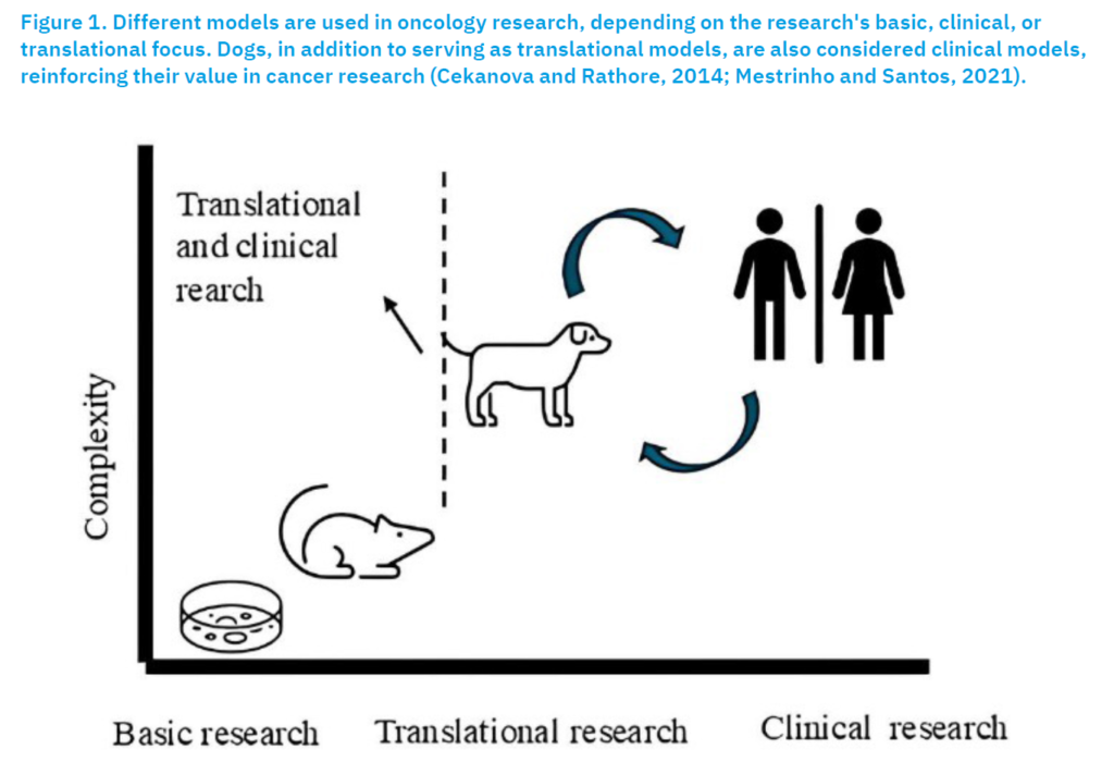
Over the past decades, companion animals, especially dogs, have gained a prominent role in comparative medicine, particularly since they develop naturally occurring tumours that reproduce the biological, genetic, and molecular characteristics observed in human neoplasms, with greater fidelity than experimentally induced models (Vail and Macewen, 2000; Fulkerson et al., 2017; Soria et al., 2019; Tarone et al., 2019). These studies have demonstrated the high translational potential of these models, as dogs share significant genetic, physiological, and clinical similarities with humans. This biological correspondence makes dogs a valuable bridge between pre-clinical research and clinical studies, benefiting both veterinary and human oncology (Figure 1).
A fundamental principle of comparative oncology is the synergistic collaboration between human and veterinary medicine. Although significant progress has been achieved, challenges remain in methodological standardisation and data integration of neoplasms (Cekanova and Rathore, 2014; Mestrinho and Santos, 2021). To address these limitations, in 2019 the United States National Cancer Institute (NCI) established the Integrated Canine Data Commons (ICDC), a digital platform designed to facilitate the sharing and analysis of genomic and clinical data from dogs with cancer, allowing direct comparisons with human neoplasms (Cekanova and Rathore, 2014; Mestrinho and Santos, 2021).
These consortia and initiatives aim to conduct clinical trials in dogs with spontaneous tumours, evaluating the pharmacokinetics and pharmacodynamics of new compounds, correlating drug exposure with modulation of tumour biomarkers, and advancing the development of more effective anticancer therapies (Cekanova and Rathore, 2014; Klingemann, 2018; Mestrinho and Santos, 2021). In parallel, systematic collection of biological samples for biobanking expands opportunities for genetic and molecular research (Cekanova and Rathore, 2014; Klingemann, 2018; Mestrinho and Santos, 2021).
The Comparative Brain Tumor Consortium and the American Veterinary Medical Association (AVMA) Animal Health Studies Database, for instance, centralise and promote clinical research in different species, including dogs with melanoma, sarcomas, osteosarcoma, and lymphoma, while also testing innovative approaches such as immunotherapy, gene therapy, and electrochemotherapy (Cekanova and Rathore, 2014; Klingemann, 2018; Mestrinho and Santos, 2021).
With advances in sequencing and genomic analysis technologies, the number of targeted therapies requiring pre-clinical validation continues to grow. However, the limited number of human patients, particularly those with rare cancers, restricts the testing of emerging treatments (Ranieri et al., 2013; Fulkerson et al., 2017; Rodrigues et al., 2023). Therefore, it is essential to use animal models that more accurately predict therapeutic outcomes in humans. In this context, dogs stand out as ideal models because they possess a functional immune system comparable to that of humans and develop spontaneous tumours that invade and metastasize naturally, enabling realistic analyses of tumour progression, therapeutic efficacy, and tumour–microenvironment interactions (Ranieri et al., 2013; Fulkerson et al., 2017; Rodrigues et al., 2023).
Dog as a Model for Studying Human Urothelial Carcinoma
Among the various types of spontaneous cancers in dogs, urothelial carcinoma (UC) is one of the most valuable models for translational research. Knapp et al. (2000) conducted an extensive study involving 102 dogs with spontaneous UC of the bladder, establishing a solid experimental foundation for investigating tumorigenic mechanisms and testing new therapeutic strategies. In contrast, spontaneous bladder cancer in mice is rare, and the development of artificial models is time-consuming, complex, and poorly reproducible, limiting their applicability (Maeda et al., 2018; Aayush et al., 2022).
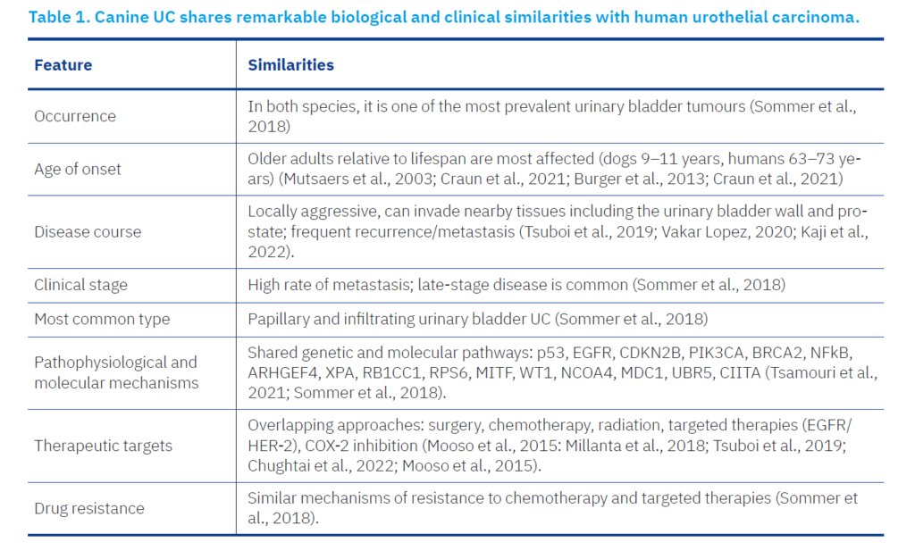
Canine UC shares remarkable biological and clinical similarities with human urothelial carcinoma, particularly in its invasive forms. Table 1 shows these biological and clinical similarities (Arnold et al., 2011; Sommer et al., 2018). Both species share homology in their proteins and genes, exhibit similar pathophysiological mechanisms of tumour initiation and progression, have comparable therapeutic targets, and show overlapping patterns of drug resistance (Arnold et al., 2011; Sommer et al., 2018).
Moreover, dogs maintain an intact immune system, enabling realistic studies of tumour: immune interactions, which are crucial for advancing immunotherapy (Arnold et al., 2011; Sommer et al., 2018).
These similarities make canine UC a natural and immunocompetent model for human muscle-invasive urothelial carcinoma, offering unique opportunities to test experimental therapies and validate diagnostic and prognostic biomarkers. One of the main clinical challenges in managing muscle-invasive UC is the lack of reliable markers to distinguish malignant from non-malignant lower urinary tract diseases (Fulkerson et al., 2017; Sommer et al., 2018; Tsamouri et al., 2024). Current studies aim to identify specific urinary biomarkers by comparing samples from dogs with muscle-invasive UC with those from dogs affected by non-malignant conditions such as urinary tract infection, urolithiasis, or proliferative urethritis (Fulkerson et al., 2017; Sommer et al., 2018; Tsamouri et al., 2024).
Because canine muscle-invasive UC occurs more frequently and spontaneously than its human counterpart, these studies are particularly advantageous, enabling faster progress in understanding tumour biology and evaluating new therapeutic approaches (Fulkerson et al., 2017; Sommer et al., 2018; Tsamouri et al., 2024). Thus, applying the principles of comparative oncology creates a proper two-way translational pathway between human and veterinary medicine, where molecular discoveries in dogs can be translated to humans, and vice versa, fostering mutual advancement in the fight against cancer (Fulkerson et al., 2017; Sommer et al., 2018; Tsamouri et al., 2024).
Epidemiology and risk factors
UC in dogs occurs predominantly in elderly animals, with an average age of 9 to 11 years, although cases in younger dogs have also been reported (Mutsaers et al., 2003; Craun et al., 2021). While dogs of all breeds can be affected, some are more predisposed, particularly Scottish Terriers, West Highland White Terriers, Wire Hair Fox Terriers, Shetland Sheepdogs, and Beagles, with the risk being particularly high in Scottish Terriers (Charney et al., 2017; Burgess and DeRegis, 2019). Other risk factors include female sex, a history of neutering, obesity, breed predisposition, and exposure to environmental chemicals (Burgess and DeRegis, 2019; Varvil and Dos Santos, 2024). Substances such as older-generation flea-control pesticides and lawn herbicides have been linked to an increased risk (Lucchesi et al., 2023).
In humans, the primary risk factor is advanced age, ranging from 63 to 73 years old (Burger et al., 2013; Craun et al., 2021). There is also a significant sex difference: men are diagnosed with bladder cancer approximately four times more often than women (Varvil and Dos Santos, 2024). This pattern may be related to differences in environmental exposures, lifestyle habits, and physiological factors, such as urinary retention secondary to prostate enlargement, which prolongs the contact of the bladder mucosa with carcinogenic agents (Burger et al., 2013; Vitti Gambim et al., 2020; Wong et al., 2023).
Interestingly, sex predisposition is reversed between species, females in dogs and males in humans, suggesting species-specific hormonal, anatomical, or metabolic modifiers of carcinogen exposure. Shared environmental factors, however, highlight dogs as sentinels for environmental bladder carcinogens that also affect humans (Knapp et al., 2014, 2020).
Histopathology
The definitive diagnosis of UC in dogs is made through histological examination of samples obtained via surgery, cystoscopy, or catheter biopsy (Inkol et al., 2021; Sakai et al., 2024). At the time of diagnosis, approximately 16% of dogs already have lymph node or distant metastases, a figure that increases to around 50% at the time of death, mainly located in the trigone area (Tsuboi et al., 2019; Vakar Lopez, 2020; Kaji et al., 2022). Urothelial carcinoma of the bladder presents distinct morphological and architectural variations, classified by the WHO (2004) in veterinary medicine as papillary or non-papillary, invasive or non-invasive tumours. Invasion can be restricted to the lamina propria or extend into the muscular layer, and in some cases, the adventitia (Knapp et al., 2014; Brambilla et al., 2022; Chen and Al-Ahmadie, 2025). In dogs, papillary and infiltrating urinary bladder UC is the most common variant, characterised by papillary projections in the bladder lumen, supported by a central fibrous stalk of variable thickness, lined by layers of neoplastic urothelium with varying degrees of atypia. The tumour cells can infiltrate the lamina propria or reach the deeper muscle layers. Non-infiltrating papillary UC, on the other hand, maintains similar luminal growth, but without stromal invasion (Knapp et al., 2014; Brambilla et al., 2022; Chen and Al-Ahmadie, 2025).
This classification also applies to human tumours. Infiltrating UCs form cohesive cell nests with amphophilic cytoplasm and large, pleomorphic, hyperchromatic nuclei that are often irregular and angular. The nucleoli vary in number and size, ranging from small to multiple or large and eosinophilic. Pleomorphism is marked by bizarre, multinucleated cells. Abnormal mitotic figures and squamous and glandular differentiation are common, with occasional mucus inclusion variations (Solomon and Hansel, 2015; Brambilla et al., 2022; Parker et al., 2024). The stroma may show desmoplasia and lymphocytic infiltration, with plasma cells. Inflammation can be mild to severe, focal or disseminated. Intraepithelial neoplasia and carcinoma in situ are frequent. Non-invasive UCs, on the other hand, have branched papillary stalks with an orderly architecture and minimal cytological variation (Solomon and Hansel, 2015; Brambilla et al., 2022; Parker et al., 2024).
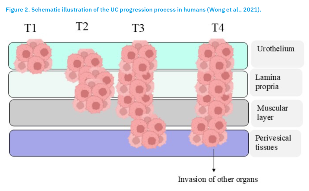
In men, invasive UC is defined as a high-grade tumour that invades the lamina propria (T1 tumours) or deeper structures, including the muscular layer (T2–T4 tumours), and is often referred to as muscle-invasive bladder cancer (Figure 2). In dogs, however, the classification is different: only stages T1, T2, and T3 are used, where T1 corresponds to invasion of the lamina propria, T2 to invasion of the muscle, and T3 to invasion of adjacent organs (Rebouissou et al., 2012; Wong et al., 2021; Pérez-Aizpurua et al., 2024; Escobar et al., 2025).
Molecular features
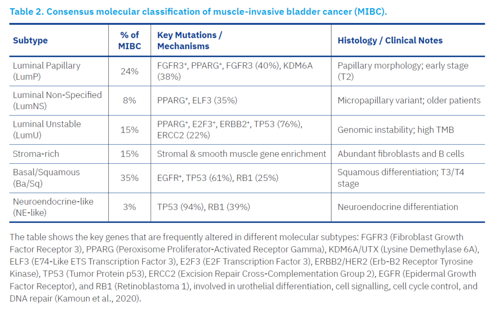
Recent molecular studies have transformed our understanding of invasive urothelial carcinoma, revealing that the disease is not homogeneous but comprises distinct gene expression based subtypes. These molecular classes parallel the basal–luminal model described initially in breast cancer, providing valuable prognostic and therapeutic insights. Importantly, invasive UC subtyping offers a framework for precision oncology, as tumour phenotype is tightly linked to aggressiveness, metastatic potential, and treatment response (McConkey and Choi, 2018; Takahara et al., 2021; Lopez-Beltran et al., 2024; Corbett et al., 2025).
Molecular subtypes
Cross-species analyses have confirmed that molecular subtypes observed in human invasive UC are also present in dogs. Transcriptomic profiling of canine tumours has identified two principal expression clusters, basal and luminal, that closely resemble the human subtypes (Choi et al., 2014; McConkey and Choi, 2018). This similarity strengthens the use of the canine model in translational studies of bladder cancer biology and therapy (Kamat et al., 2016; Chen and Al-Ahmadie, 2025).
Luminal tumours typically exhibit papillary histoarchitecture and are associated with a favourable outcome (Choi et al., 2014; Kamat et al., 2016). Their molecular signature includes the expression of HER, TRIM24, FOXA1, GATA3, and PPARG, as well as frequent FGFR3 mutations, which serve as predictive markers of response to FGFR inhibitors (Table 2). These tumours respond better to certain targeted therapies than basal variants (Cheng et al., 2012; Rebouissou et al., 2012; Choi et al., 2014)
Basal invasive UCs, in contrast, show enrichment of squamous differentiation and are often diagnosed in more advanced stages (Choi et al., 2014; Kamat et al., 2016; Kamoun et al., 2020). They are particularly common in females and present a highly aggressive clinical course. The basal transcriptomic profile includes upregulation of STAT3, TP63, KRT5/6A, and CD44, as well as activation of the NFκB, c-Myc, and HIF pathways. Some tumours also display epithelial-to-mesenchymal transition markers, aligning with “claudin-low” breast cancer signatures. Despite their aggressiveness, these tumours may be more responsive to platinum-based chemotherapy and immune checkpoint inhibitors (Choi et al., 2014; Al-Ahmadie and Iyer, 2018; Sarfaty et al., 2023) (Table 2).
Other Molecular Pathways in Invasive UC
Although the genomic landscape of canine invasive urothelial carcinoma is still being elucidated, current evidence reveals remarkable similarities with human tumours across several key oncogenic pathways.
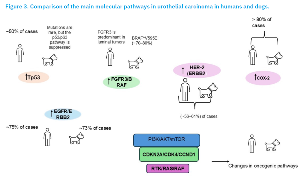
Mutations in TP53 are among the most frequent in human invasive urothelial carcinoma (InvUC), occurring in approximately half of all cases (Mojarrad and Moghbeli, 2020). These often coexist with RB1 deletions and MDM2 amplification, collectively disabling major tumour suppressor functions. Although direct TP53 mutations are rare in canine InvUC, transcriptomic analyses demonstrate suppression of p53 regulatory networks and reduced p63 expression, indicating a similar functional impairment of the p53/p63 axis (Damrauer et al., 2014; Czerniak, 2016; Varchulová Nováková et al., 2023). Aberrant signalling contributes to tumour growth in approximately 40% of human InvUCs (Knowles, 2008; Mojarrad and Moghbeli, 2020). Overexpression of this pathway’s components has also been observed in dogs, suggesting conserved oncogenic activation. Ongoing studies are assessing this cascade as a shared therapeutic target across species (Rouanne et al., 2016).
EGFR overexpression is seen in approximately 75% of human and 73% of canine high-grade tumours. Although EGFR inhibitors show variable efficacy, they tend to yield better results in chemotherapy-naïve patients or in tumours with ERBB2/EGFR amplification (Mooso et al., 2015; Soria et al., 2019b; Vitti Gambim et al., 2020; Ozsagir et al., 2024). Experimental therapies, such as photoimmunotherapy (Can225-IR700) and EGFR-targeted toxins, have shown promising pre-clinical results in dogs (Raghavan, 2015; Sakai et al., 2020, 2024).
While FGFR3 mutations predominate in human luminal tumours and BRAF alterations are uncommon, canine InvUC is characterised by the BRAFV595E mutation, analogous to BRAFV600E in humans, detected in roughly 70–80% of cases (Rouanne et al., 2016; Kaji et al., 2022; Guercio et al., 2023; Loriot et al., 2023; Aeschlimann et al., 2024). This mutation leads to constitutive activation of the MAPK pathway, illustrating a divergence in initiating events but a convergence in downstream proliferative signalling dysregulation across species (Soria et al., 2019).
Similarly, HER-2 (ERBB2) is detected in approximately 56–61% of canine InvUCs, a proportion comparable to that observed in human cases. This cross-species expression pattern supports the development of HER-2, targeted therapies in translational oncology (Laé et al., 2010; Millanta et al., 2018; Tsuboi et al., 2019; Chughtai et al., 2022).
COX-2 (cyclooxygenase-2) is another marker consistently overexpressed in more than 80% of invasive urothelial carcinoma cases in both species, as shown by immunohistochemical and transcriptomic analyses (Yildirim et al., 2010; Al-Maghrabi et al., 2019). This observation reinforces the role of inflammation in tumorigenesis and explains the clinical benefit of nonsteroidal anti-inflammatory drugs (NSAIDs), such as piroxicam, in treating canine invasive UC (Gordon et al., 2009; Gardner et al., 2016; Knapp et al., 2016; Ciriano Cerdà et al., 2023).
High-throughput sequencing studies have revealed extensive overlap in the genomic alterations of human and canine invasive urothelial carcinomas, including recurrent disruptions in the PI3K/AKT/mTOR, CDKN2A/CDK4/CCND1, and RTK/RAS signalling pathways (Figure 3) (Rouanne et al., 2016; Korec et al., 2021; Dawid De Vera et al., 2023).
In canine invasive UC, over 1500 genes are differentially expressed compared with normal bladder tissue, with approximately 450 genes dysregulated shared with the human counterpart (Maeda et al., 2018). Notable examples include CDKN2B, PIK3CA, BRCA2, NFκB, ARHGEF4, MDC1, RB1CC1, MITF, and WT1, reflecting both conserved and species-specific oncogenic mechanisms (Rouanne et al., 2016; Korec et al., 2021; Dawid De Vera et al., 2023) (Figure 3).
The standard treatment for human invasive urothelial carcinoma involves cystectomy to control the primary tumour and systemic chemotherapy to manage metastases. Radiotherapy may be employed as an alternative for bladder preservation or regional disease control. As in dogs, approximately 50% of human patients develop metastases to regional lymph nodes, lungs, and other organs, these being the leading cause of mortality (Wong et al., 2023; Varvil and Dos Santos, 2024). Anatomically, however, the tumour tends to arise at the bladder apex in humans, whereas in dogs it more commonly occurs at the bladder neck (Wong et al., 2023; Varvil and Dos Santos, 2024).
Numerous molecular alterations are associated with urothelial carcinoma. Frequent mutations include FGFR3, PIK3CA, STAG2, and genes in the RTK/RAS/RAF pathway in non–muscle-invasive bladder cancers (NMIBCs), whereas ERBB2, RB1, MDM2, TP53, CDKN2A, ARID1A, and KDM6A predominate in muscle-invasive bladder cancers (MIBCs). Among these, TERT promoter mutations are particularly recurrent and play a crucial role in tumorigenesis (Raghavan, 2015; Sweis and Galsky, 2016; Soria et al., 2019; Yoshitake et al., 2020; Schwarzova et al., 2023).
From an immunological perspective, several T-cell modulators are implicated in urothelial carcinoma. Co-stimulatory molecules such as 4-1BB (CD137), OX-40 (CD134), GITR, and ICOS (CD278) promote T-cell activation and proliferation, enhancing the antitumor immune response (Rouanne et al., 2016; Sweis and Galsky, 2016; Knapp et al., 2020; Schwarzova et al., 2023). Conversely, inhibitory molecules like CTLA-4 (CD152), LAG-3 (CD223), Tim-3, VISTA, BTLA-4, and PD-1 (CD279) negatively regulate immune activity, facilitating tumour immune evasion and contributing to T-cell dysfunction and tolerance within the tumour microenvironment (Rouanne et al., 2016; Sweis and Galsky, 2016; Knapp et al., 2020; Schwarzova et al., 2023).
The most widely used marker of urothelial differentiation in dogs is uroplakin III. In addition, COX-2 is overexpressed in both invasive transitional cell carcinoma (TCC) and carcinoma in situ, but not in the normal urothelium, reinforcing its potential as a diagnostic and therapeutic biomarker in both species (Sweis and Galsky, 2016; Schwarzova et al., 2023).
Conclusion and future remarks
Canine urothelial carcinoma has increasingly been recognised as a valuable spontaneous model for the study of human bladder cancer. The close resemblance between the two species, spanning histopathological features, tumour progression patterns, molecular alterations, and therapeutic responses, reinforces the translational relevance of the canine disease. Unlike rodent models, which require artificial tumour induction, dogs develop UC naturally, within an intact immune system and under environmental conditions comparable to those experienced by humans. This provides a biologically realistic setting for investigating tumour initiation, invasion, metastasis, and treatment outcomes.
Molecular studies further highlight this alignment. The identification of basal and luminal phenotypes in canine tumours, together with dysregulation of key pathways such as EGFR, PI3K/AKT/mTOR, MAPK, and COX-2, mirrors patterns well documented in human UC. Clinically relevant mutations, including the BRAFV595E variant common in dogs, highlight both shared mechanisms of tumorigenesis and species-specific distinctions, advancing a more comprehensive understanding of the disease. The naturally high frequency of invasive UC in dogs also offers an opportunity to assess emerging therapies, immunomodulatory approaches, and novel biomarkers with greater translational impact.
Continued progress in this field will depend heavily on collaborative efforts that integrate clinical, genomic, and therapeutic data across institutions. Initiatives such as the Integrated Canine Data Commons have begun to support these objectives. Still, broader adoption, methodological standardization, and expansion of clinical trials in dogs with spontaneous tumours will be essential. Likewise, investment in tissue and biofluid biobanking, combined with advances in sequencing technologies, will facilitate more profound exploration of tumour heterogeneity, progression pathways, and mechanisms of therapeutic resistance.
Funding: This work was carried out within the scope of the AICanceR∞Hub project (NORTE2030-FEDER-02687800), co-financed by the European Regional Development Fund (ERDF) under the Northern Regional Program 2021-2027 [NORTE2030].
Acknowledgments: Project UIDB/00772/2020 funded by the Portuguese Foundation for Science and Technology (FCT).
References [… show]
Urotelijalni karcinom kod pasa: obećavajući model za napredak istraživanja humanog urotelijalnog karcinoma?
Ana Rita TEIXEIRA FILES*, (dopisni autor), ritafiles2000@gmail.com, orcid.org/0009-0009-6211-850X; Isabel Cristina RIBEIRO PIRES, ipires@utad.pt, orcid.org/0000-0001-6330-4560.
University of Tras-os-Montes and Alto Douro, 5000-801 Vila Real, Portugal
Psi su vrijedni modeli u komparativnoj onkologiji jer spontano razvijaju tumore, imaju kraći životni vijek i dijele značajne biološke i kliničke sličnosti s humanim tumorima. Cilj ovog rada je procijeniti relevantnost psećeg urotelijalnog karcinoma (UC) kao translacijskog modela za ljudski rak mokraćnog mjehura usporedbom epidemioloških obrazaca, molekularnih promjena i imunoloških značajki među vrstama. Urotelijalni karcinom je od posebnog interesa zbog svoje agresivne prirode i visokih stopa smrtnosti. Kod pasa su invazivni oblici UC-a češći, što naglašava relevantnost ove vrste za modeliranje ljudskih bolesti. Iako su starija dob i izloženost kancerogenima iz okoliša zajednički čimbenici rizika, postoje značajne epidemiološke razlike, uključujući veću prevalenciju kod ženki pasa nego kod muškaraca. Molekularno, pseći i ljudski UC dijele podtipove tumora i disregulaciju ključnih onkogenih putova, kao što su p53/p63, RTK/RAS/MAPK, PI3K/AKT/mTOR i CDKN2A/CDK4, unatoč različitim inicijacijskim genetskim događajima. Ove promjene konvergiraju u putovima koji kontroliraju proliferaciju, preživljavanje i izbjegavanje imunološkog odgovora. Zajednička prekomjerna ekspresija terapijskih ciljeva i slični mehanizmi imunoloških kontrolnih točaka dodatno podupiru translacijsku vrijednost psećeg modela. Sve ove značajke pojačavaju korisnost pasa u unapređenju biološkog razumijevanja i terapijskog razvoja za urotelijalni karcinom u veterinarskoj i humanoj medicini.
Ključne riječi: urotelijalni karcinom; tranzicijski karcinom; translacijski modeli; komparativna onkologija; pas.