
Microbiological Safety Assessment of Canine Adipose Derived Mesenchymal Stem Cell Preparations Reveal no Detectable Contamination
M. Prišlin Šimac , V. Kunić*, D. Brnić, D. Jurković Žilić, R. Beck, S. Duvnjak, I. Reil, M. Zadravec, V. Kostanić
Marina PRIŠLIN ŠIMAC1, prislin@veinst.hr, orcid.org/0000-0002-1169-2060; Valentina KUNIĆ2*(corresponding author), kunic@veinst.hr, orcid.org/0000-0002-0154-7652; Dragan BRNIĆ2, brnic@veinst.hr, orcid.org/0000-0002-7318-8337; Daria JURKOVIĆ ŽILIĆ3, jurkovic@veinst.hr, orcid.org/0000-0002-3504-5271; Relja BECK3, beck@veinst.hr, orcid.org/0000-0002-5304-2875; Sanja DUVNJAK4, marjanovic@veinst.hr, orcid.org/0000-0002-1308-267X; Irena REIL4, reil@veinst.hr, orcid.org/0000-0002-2198-557X; Manuela ZADRAVEC5, zadravec@veinst.hr; orcid.org/0000-0003-4382-4424; Vjekoslava KOSTANIĆ2, kostanic@veinst.hr, orcid.org/0009-0009-6720-1329.
1Laboratory for Rabies and General Virology, Department for Virology, Croatian Veterinary Institute, 10000 Zagreb, Croatia
2Laboratory for Serological Diagnosis of Viral Diseases, Department for Virology, Croatian Veterinary Institute, 10000 Zagreb, Croatia
3Laboratory for Parasitology, Department for Bacteriology and Parasitology, Croatian Veterinary Institute, 10000 Zagreb, Croatia
4Laboratory for Bacterial Zoonoses and Molecular Diagnosis of Bacterial Diseases, Department for Bacteriology and Parasitology, Croatian Veterinary Institute, 10000 Zagreb, Croatia
5Laboratory for Feed Microbiology, Department for Veterinary Public Health, Croatian Veterinary Institute, 10000 Zagreb, Croatia

https://doi.org/10.46419/cvj.57.6.10
Abstract
The experimental application of canine adipose-derived mesenchymal stem/stromal cells (cAD-MSC) has demonstrated excellent therapeutic potential in the treatment of a number of previously untreatable or difficult-to-treat diseases. However, the production of cAD-MSC for clinical application necessitates in vitro isolation and cell expansion, introducing the risk of microbiological contamination. Studies indicate that stored MSCs may be contaminated with bacteria, fungi, or viruses, resulting in cell transformation, cell death, or persistent infection. This ultimately compromises therapeutic efficacy and potentially facilitates therapy-induced pathogen transmission. In accordance with the European Medicines Agency guidelines mandating sterility testing as an integral component of the manufacturing process, this study aimed to assess the sterility of the cAD-MSC bank at the Croatian Veterinary Institute. To test for the most clinically significant canine pathogens, nucleic acids were extracted from 32 cAD-MSC preparations and analyzed using standard molecular PCR methods. The pathogen screening panel included rabies virus (Lyssavirus), canine herpesvirus (Varicellovirus canidalpha1), parvovirus (Carnivore protoparvovirus 1), coronavirus (Alphacoronavirus 1), distemper virus (Morbillivirus canis), Rotavirus A, Leptospira spp, Brucella canis, Borrelia burgdorferi sensu lato, Anaplasma spp, Ehrlichia spp, Mycoplasma spp (hemotropic), and Neospora caninum. No tested gene fragments of the aforementioned pathogens were detected, indicating that the cAD-MSC preparations were free from the presence of clinically significant pathogens. Rigorous implementation of sterility control during the cell isolation and expansion process is essential to ensure safe cell-based therapy in canine patients.
Key words: adipose-derived mesenchymal stem cells; regenerative veterinary medicine; cell therapy; dog; microbial contamination.
Introduction
Canine adipose-derived mesenchymal stem/stromal cells (cAD-MSC) have emerged as a promising therapeutic option in veterinary medicine, particularly in the treatment of canine degenerative diseases previously considered untreatable or challenging to manage (Voga et al., 2020). Experimental applications of cAD-MSC have demonstrated remarkable results across a range of conditions, including orthopedic, neurological, dermatological, ophthalmological, gastroenterological, and hematological pathologies (Prišlin et al., 2022). These successes highlight the therapeutic potential of cAD-MSC and their capacity to improve clinical outcomes in canine medicine. The production of cAD-MSC for therapeutic purposes necessitates in vitro isolation and subsequent cell expansion, processes that inherently carry the risk of microbiological contamination. Contamination can occur at various stages of cell processing and poses a significant threat to the safety and efficacy of MSC-based therapies. Studies have reported contamination rates as high as 40% in stored MSC preparations, with several types of bacteria, moulds/yeasts, and viruses (Martín et al., 2012; Pekker et al., 2023; Szabłowska-Gadomska et al., 2023). Such contamination can lead to serious consequences, including cell transformation, cell death, or persistent infections (Khatri et al., 2010; Nazari-Shafti et al., 2011; Jacobs et al., 2013; Khatri and Saif, 2013). These adverse outcomes not only compromise the viability and functionality of MSCs but could also result in therapy failure or the transmission of pathogens to treated animals, thereby undermining the therapeutic benefits of cAD-MSC.
To ensure the safety and quality of MSC preparations, rigorous sterility testing is essential. The European Medicines Agency emphasises that sterility testing is a mandatory component of the manufacturing process for cell-based therapeutic products (Anonymous, 2016). Adherence to these guidelines is critical for minimising the risks associated with microbiological contamination and for safeguarding the therapeutic efficacy of MSC preparations. In line with these requirements, the present study focused on evaluating the sterility of the cAD-MSC bank maintained by the Croatian Veterinary Institute. The primary aim was to apply standard sterility testing of cell culture with broths and assess the presence of specific, most significant pathogens, including rabies virus (Lyssavirus), canine herpesvirus (Varicellovirus canidalpha1; CHV), parvovirus (Carnivore protoparvovirus 1; CPV), coronavirus (Alphacoronavirus 1; CCoV), distemper virus (Morbillivirus canis; CDV), Rotavirus A (RVA), Leptospira spp, Brucella canis, Borrelia burgdorferi sensu lato, Anaplasma spp, Ehrlichia spp, Mycoplasma spp (hemotropic), and Neospora caninum. These pathogens represent a significant health risks to dogs, representing an important step in ensuring the microbiological quality of cAD-MSC. This research aligns with global efforts to advance the safe application of stem cell preparations in regenerative veterinary medicine.
Material and Methods
Extraction, storage and characterization of cAD-MSC
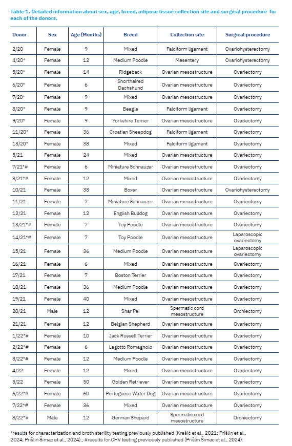
Adipose tissue samples were collected from 32 clinically healthy dogs (Canis lupus familiaris; 30 males, 2 females), presenting for elective surgical procedures. Table 1 provides detailed information regarding the age, breed, adipose tissue collection site, and tissue mass for each donor. The procedures for adipose tissue collection, isolation of cAD-MSC, and their subsequent propagation were conducted following previously established protocols (Krešić et al., 2021). All donor cells were cryopreserved in liquid nitrogen using the standard cryobanking protocol with 10% dimethyl sulfoxide (Sigma-Aldrich, St. Louis, MO, USA; Cat. No. D2650-100ML) at passage 2 (P2) or P3 as described previously (Prišlin Šimac et al., 2024). The extracted cAD-MSCs were characterized by immunophenotyping and three-lineage differentiation to conform to the criteria established by the International Society for Cellular Therapy (Dominici et al., 2006), results were previously published for all donors marked with asterisks in Table 1 (Krešić et al., 2021; Prišlin et al., 2023, 2024; Prišlin Šimac et al., 2024).
Sterility assessment of cAD-MSC Cultures
Sterility assessment was performed immediately after the extraction of cAD-MSCs from adipose tissue, using in-house broth-based methods for detecting aerobic bacteria, anaerobic bacteria, and moulds/yeasts Moreover, tryptic soy broth with casein tested for aerobic bacteria, thioglycolate broth with resazurin for anaerobic bacteria, and 2% Sabouraud broth for moulds/yeasts. All broths were incubated at 37°C under aerobic conditions, except for thioglycolate, which was maintained under anaerobic conditions. The results of standard sterility testing for all donors marked with asterisks in Table 1 have been previously published (Krešić et al., 2021; Prišlin et al., 2024; Prišlin Šimac et al., 2024).
Extraction of total nucleic acids of cAD-MSC Cultures
A total of 32 cAD-MSC donor samples stored in the Croatian Veterinary Institute biobank were subjected to total nucleic acid extraction. One cryobanked batch of cells per donor at P2 or P3 was first transferred at -20°C to induce lysis of the cell membranes. After 24 h, the cell lysate was thawed at room temperature for 30 min, vortexed and subjected to nucleic acid extraction using a MagMAX CORE nucleic acid purification kit (Thermo Fisher Scientific, Waltham, USA) on the KingFisher Flex Purification System (Thermo Scientific), in accordance with the manufacturer’s instructions. Before conducting subsequent polymerase chain reaction (PCR)-based detection of the targeted pathogens, the success of nucleic acid extraction for each sample was verified by PCR amplification of an endogenous internal control, the β-actin gene, as described by Wernike et al. (2011).
Detection of viral pathogens
Canine Herpes Virus and Canine Parvovirus
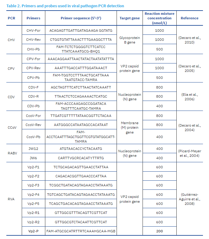
Target regions for CHV and CPV real-time PCR detection included the glycoprotein B (gB) gene (Decaro et al., 2010) and the VP2 capsid protein gene (Decaro et al., 2005), respectively. Amplification was conducted using the QuantiFast Pathogen PCR+IC kit (Qiagen, Hilden, Germany) on the Rotor-Gene Q system (Qiagen) following the manufacturer’s cycling conditions. Results of sterility testing for CHV were previously published for donors marked with the hash sign in Table 1. Primer and probe details are provided in Table 2.
Canine Distemper Virus and Canine Coronavirus
Real-time reverse transcription PCR (RT-PCR) targeted the nucleoprotein (N) gene (Elia et al., 2006) and the membrane protein (M) gene encoded in ORF5 (Decaro et al., 2004) for CDV and CCoV, respectively. Target regions were amplified using the AgPath One-Step RT-PCR kit (Applied Biosystems, Foster City, USA) on the Rotor-Gene Q system (Qiagen). The assay was performed according to the manufacturer’s instructions, with a modified initial reverse transcription step for 10 minutes at 50°C for the CDV. Primer and probe details are specified in Table 2.
Rabies Virus
For RABV, conventional RT-PCR targeted the nucleoprotein (N) gene (Picard-Meyer et al., 2004), using the SuperScript III One-Step RT-PCR System with Platinum Taq polymerase kit (Thermo Fisher Scientific) on the 2720 Thermal Cycler (Applied Biosystems). The amplification was performed with the initial RT step for 30 minutes at 50°C, 2 minutes at 94°C, followed by 35 cycles of 45 seconds at 94°C, 45 seconds at 48°C and for 1 minute at 68°C. Product visualization was performed via capillary electrophoresis on the QIAxcel Advanced system using the DNA Screening Kit and DNA Size Marker 100bp–2,5kb (Qiagen) as per the manufacturer’s instructions. Primer details are specified in Table 2, while expected amplicon size followed referenced protocol.
Rotavirus A
For RVA, real-time RT-PCR targeted the VP2 capsid protein gene (Gutiérrez-Aguirre et al., 2008) with the VetMAX-Plus One-Step RT-PCR Kit (Applied Biosystems). Prior to amplification, RNA was combined with primers and subjected to a denaturation step for 5 minutes at 95°C. Amplification was performed on the Rotor-Gene Q system (Qiagen) following the manufacturer’s cycling conditions for MGB probes. Primer and probe details are specified in Table 2.
Detection of Bacterial and Parasitic Pathogens
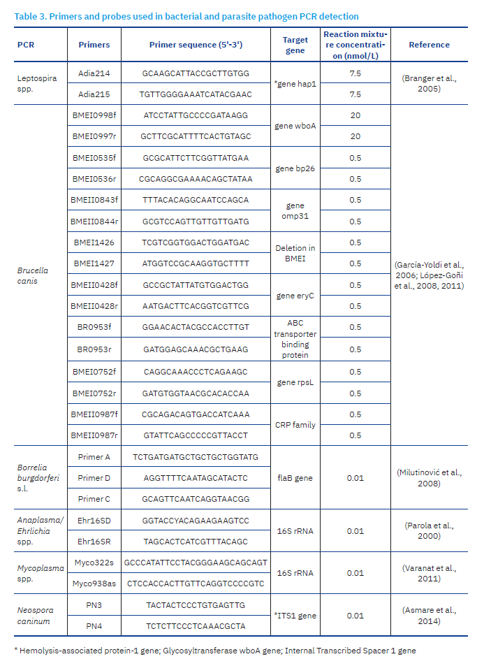
For each bacterial and parasitic pathogen described in this section, PCR cycling was conducted on the SimpliAmp Thermal Cycler (Thermo Fisher Scientific), followed by amplicon visualization as described earlier for RABV. Primer details for each pathogen in this section are specified in Table 3, while the expected amplicon sizes followed referenced protocols.
Leptospira spp., Brucella canis, Borrelia burgdorferi sensu lato and Mycoplasma spp.
Leptospira spp. detection was based on conventional PCR targeting the hemolysis-associated protein-1 gene (hap1), performed as previously described (Branger et al., 2005) with HotStarTaq Master Mix Kit (Qiagen).
B.canis was screened using the Bruce-ladder multiplex PCR assay (García-Yoldi et al., 2006; López-Goñi et al., 2008, 2011), with Multiplex PCR Kit (Qiagen). PCR amplification consisted of an initial denaturation at 95°C for 15 minutes, followed by 35 cycles of 30 seconds at 95°C, 45 seconds at 64°C and 3 minutes at 72°C, with a final extension at 72°C for 10 minutes.
Borrelia burgdorferi sensu lato was screened by semi-nested PCR targeting the flaB gene according to Milutinović et al. (2008), while Mycoplasma spp. detection targeted the 16S rRNA gene as described by Varanat et al. (2011). Reactions were performed using the GoTaq G2 Master Mix (Promega, Madison, USA), following the cycling conditions described in referenced protocols.
Anaplasma/Ehrlichia spp. and Neospora caninum
All samples were further screened for Anaplasma/Ehrlichia species using PCR assay amplifying a fragment of the 16S rRNA gene, as previously described (Parola et al., 2000). Detection of Neospora caninum was performed using a PCR assay targeting the Internal Transcribed Spacer 1 gene (ITS1) region, as previously described by Asmare et al., 2014. Both reactions were performed using the GoTaq G2 Master Mix (Promega), following the cycling conditions and amplicon characteristics described in the referenced protocol.
Results
Isolation, storage and characterization of cAD-MSC immunophenotype and differentiation potential
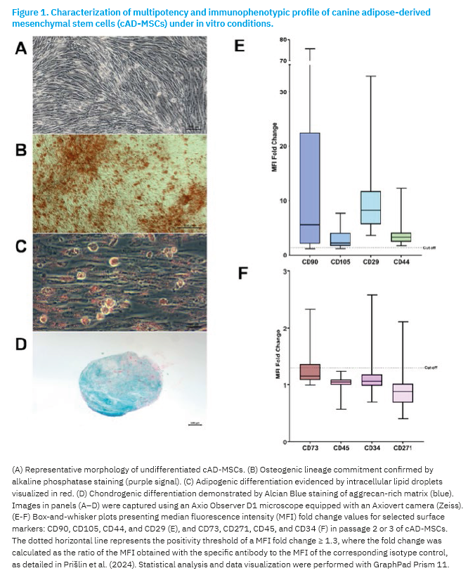
All 32 cAD-MSCs were successfully isolated from adipose tissue. Cultures exhibited the expected spindle-shaped morphology (Figure 1A), along with gradual cellular enlargement during prolonged in vitro cultivation. cAD-MSCs from all donors demonstrated trilineage differentiation capacity, as evidenced by successful induction into adipogenic (red lipid droplets), osteogenic (red calcium deposits), and chondrogenic (turquoise aggrecan-positive) lineages (Figures 1B-D). Furthermore, flow-cytometric analysis of cells at P2 or P3 confirmed consistent expression of cluster of differentiation (CD) 90, CD29, CD105, and CD44 (Figure 1E) in all donors. CD45, CD271, CD34 were not detected, while CD73 showed low-level expression (Figure 1F).
Sterility assessment of cAD-MSC cultures by broths
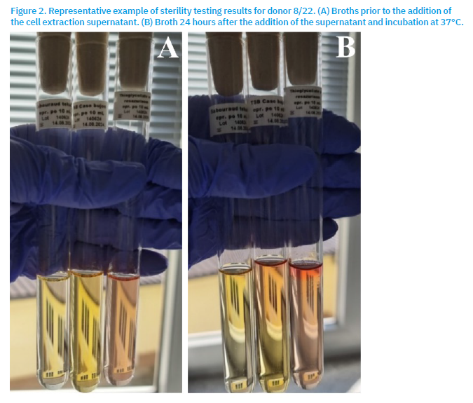
Sterility testing demonstrated the absence of microbial contamination in all cAD-MSC samples during extraction. A representative example of these sterility testing results is shown in Figure 2.
Pathogen screening
PCR analysis consistently detected β-actin in all samples (Cq ~24–28), confirming that an adequate number of cells were present and efficiently lysed for downstream molecular analyses. Amplification of β-actin also verified the absence of PCR inhibitors, supporting the validity of all subsequent pathogen-specific assays.
Comprehensive molecular screening revealed no detectable tested gene fragments of any of the targeted pathogens, including CHV, CPV, CCoV, CDV, RVA, Leptospira spp, Brucella canis, Borrelia burgdorferi sensu lato, Anaplasma spp, Ehrlichia spp, Mycoplasma spp (hemotropic), and Neospora caninum. All negative results were validated by appropriate positive and negative assay controls. Together, these findings indicate that cAD-MSCs used in this study were not contaminated with any of the tested pathogens.
Discussion
In the present study, we assessed microbial contamination in cAD-MSC preparations from the cAD-MSC bank maintained at the Croatian Veterinary Institute. We focused on cell culture sterility testing using broths for the detection of bacterial and fungal contamination, and various PCR methods for the molecular detection of clinically significant canine viral, bacterial and parasitic pathogens. The tested pathogens are clinically significant and represent a substantial health risks to dogs, hence screening for their presence is a critical step in ensuring the microbiological safety and therapeutic suitability of cAD-MSC preparations. The results demonstrated that none of the cAD-MSC preparations were contaminated, in particular with any of the tested pathogens, as also seen in Luo et al. (2021). This finding suggests that the cAD-MSC bank maintained at the Croatian Veterinary Institute is free of tested microbial contaminants, which is a crucial prerequisite for the safe clinical application of these cells. In particular, the absence of Mycoplasma spp. is highly relevant, as mycoplasmas are among the most frequent and problematic contaminants in cell culture. Due to their small size, lack of a rigid cell wall, and slow growth, mycoplasmas often escape routine microscopic detection, yet they can profoundly alter cell metabolism, proliferation, gene expression, membrane antigenicity, and differentiation capacity, thereby compromising both experimental reproducibility and therapeutic safety (Mahmood and Ali, 2017; Wilder and Reid, 2022).
While the absence of investigated pathogens in the tested cAD-MSC preparations is a positive outcome, it must be emphasized that the risk of contamination can never be eliminated. Previously reported viral contaminants in human cell cultures include viruses including hepatitis viruses, retroviruses, herpes viruses or papillomaviruses, whereas contamination events in animal cell lines are documented less frequently (Mahmood and Ali, 2017), likely reflecting the lower intensity of surveillance and reporting compared to human biomedical research. Nevertheless, animal cell cultures are inherently prone to contamination because they are maintained in nutrient-rich media that support rapid microbial growth, and because primary cultures often originate from tissues with endogenous microbiota or latent infections (Mahmood and Ali, 2017). Viral contamination remains significantly more challenging (Merten, 2002; Niehues et al., 2020) than bacterial and fungal contamination, considering those can be partially controlled using antibiotics and antifungals. Nevertheless, the more recent opinion discourages the routine use of antibiotics/antimycotics in stem-cell banking, as they may mask low-grade contamination and promote the emergence of resistant microorganisms (Mahmood and Ali, 2017). Instead, sterility should be ensured primarily through strict adherence to Good Laboratory Practice, and the implementation of robust sterility control measures.
During routine handling of cell cultures, several indicators of potential microbial contamination should be carefully monitored, including unexpected changes in culture medium turbidity or color, altered pH, unusual odors, reduced cell adherence, slowed or accelerated proliferation, abnormal cell morphology, increased cell debris, vacuolization, or spontaneous detachment (Herman and Pauwels, 2014; Weiskirchen et al., 2023). Importantly, some contaminants such as mycoplasma or latent viral infections may not produce obvious visual changes, reinforcing the need for scheduled molecular and microbiological testing (Mahmood and Ali, 2017; Feng et al., 2019). Regular testing for a comprehensive panel of pathogens, as performed in this study, can help ensure the safety and quality of cAD-MSC preparations intended for clinical use.
The clinical relevance of stringent sterility testing is underscored by reports of cases in which contaminated stem cell products were administered to patients. However, infectious complications immediately following the infusion of autologous or allogeneic contaminated grafts are uncommon (Damonti et al., 2021), and major adverse outcomes have rarely been reported (Klein et al., 2006). The microbial contamination of autologous stem cell products was concluded a rare event, affecting only 0.6% of all products as described in Damonti et al. (2021). Regardless of rarity, clinical cases emphasize the ethical and regulatory responsibility of cell banks to implement robust biosafety screening prior to clinical application.
Collectively, the presented results demonstrate that the validated molecular assays provide a reliable, sensitive, and comprehensive platform for biosafety assessment of canine MSC preparations. Their implementation supports the establishment of standardized quality control frameworks for veterinary regenerative medicine, significantly reducing the risk of transmitting infectious agents to animal patients.
Acknowglements
The authors acknowledge the support of the Croatian Science Foundation in the preparation of this research article, provided through funding under the Installation Research Project (UIP-2019-04-2178) “Revealing the Mesenchymal Stem Cells Transcriptome and Secretome” – SECRET. The authors would also like to thank Ivana Ljolje and Petar Kostešić for adipose tissue sourcing, and Mihaela Stuparić Komušar, Dunja Vlahović and Šimun Naletilić for their help with spheroid preparation and detection.
References [… show]
Procjena prisutnosti patogena u pripravcima mezenhimalnih matičnih stanica masnog tkiva pasa
Marina PRIŠLIN ŠIMAC1, prislin@veinst.hr, orcid.org/0000-0002-1169-2060; Valentina KUNIĆ2*(dopisni autor), kunic@veinst.hr, orcid.org/0000-0002-0154-7652; Dragan BRNIĆ2, brnic@veinst.hr, orcid.org/0000-0002-7318-8337; Daria JURKOVIĆ ŽILIĆ3, jurkovic@veinst.hr, orcid.org/0000-0002-3504-5271; Relja BECK3, beck@veinst.hr, orcid.org/0000-0002-5304-2875; Sanja DUVNJAK4, marjanovic@veinst.hr, orcid.org/0000-0002-1308-267X; Irena REIL4, reil@veinst.hr, orcid.org/0000-0002-2198-557X; Manuela ZADRAVEC5, zadravec@veinst.hr; orcid.org/0000-0003-4382-4424; Vjekoslava KOSTANIĆ2, kostanic@veinst.hr, orcid.org/0009-0009-6720-1329.
1Laboratorij za bjesnoću i opću virologiju, Odjel za virologiju, Hrvatski veterinarski institut, 10000 Zagreb, Hrvatska
2Laboratorij za serološku dijagnostiku virusnih bolesti, Odjel za virologiju, Hrvatski veterinarski institut, 10000 Zagreb, Hrvatska
3Laboratorij za parazitologiju, Odjel za bakteriologiju i parazitologiju, Hrvatski veterinarski institut, 10000 Zagreb, Hrvatska
4Laboratorij za bakterijske zoonoze i molekularnu dijagnostiku bakterijskih bolesti, Odjel za bakteriologiju i parazitologiju, Hrvatski veterinarski institut, 10000 Zagreb, Hrvatska
5Laboratorij za mikrobiologiju hrane za životinje, Odjel za veterinarsko javno zdravstvo, Hrvatski veterinarski institut, 10000 Zagreb, Hrvatska
Sažetak
Eksperimentalna primjena mezenhimskih matičnih stanica porijeklom iz masnog tkiva pasa (cAD-MSC) pokazala je izniman terapijski potencijal u liječenju različitih bolesti koje su prethodno bile neizlječive ili teško izlječive. Međutim, tijekom proizvodnje cAD-MSC pripravaka za terapijsku primjenu, nužno je in vitro izdvajanje i umnažanje stanica, što uvodi rizik mikrobiološke kontaminacije. Istraživanja ukazuju na to da pohranjene MSC mogu biti kontaminirane bakterijama, kvascima/plijesnima ili virusima, što može rezultirati transformacijom stanica, staničnom smrću ili perzistentnom infekcijom. To u konačnici narušava terapijsku učinkovitost i potencijalno omogućuje prijenos patogena putem terapije. Sukladno smjernicama Europske agencije za lijekove, koje nalažu provođenje ispitivanja sterilnosti kao sastavnog dijela proizvodnog procesa, cilj ovog istraživanja bio je procijeniti sterilnost banke cAD-MSC pri Hrvatskom veterinarskom institutu. U svrhu detekcije klinički najznačajnijih patogena kod pasa, iz 32 pripravka cAD-MSC izolirane su nukleinske kiseline te analizirane standardnim molekularnim PCR metodama. Panel patogena uključivao je pseći herpesvirus (Varicellovirus canidalpha1), parvovirus (Carnivore protoparvovirus 1), koronavirus (Alphacoronavirus 1), virus štenećaka (Morbillivirus canis), Rotavirus A, Leptospira spp., Brucella canis, Borrelia burgdorferi sensu lato, Anaplasma spp., Ehrlichia spp., Mycoplasma spp. (hemotropne) i Neospora caninum. Primjenom navedenih metoda nije utvrđena prisutnost odsječaka genoma navedenih patogena, odnosno nije utvrđena mikrobna kontaminacija pripravaka cAD-MSC. Unatoč tome, stroga provedba kontrole sterilnosti tijekom procesa izolacije i umnožavanja stanica nužna je kako bi se osigurala sigurna primjena stanične terapije u pasa.
Ključne riječi: mezenhimske matične stanice iz masnog tkiva; regenerativna veterinarska medicina; stanična terapija; pas; mikrobiološka kontaminacija.
