
Pelvic Fractures in Dogs: Surgical Treatment Options and Postoperative Management
M.Drašić, N. Ivkić, M. Pećin, P. Kostešić*, S. Faraguna
Martina DRAŠIĆ1, drasicmartina@gmail.com; Niko IVKIĆ2, nivkic@vef.unizg.hr; Marko PEĆIN2, mpecin@vef.unizg.hr, orcid.org/0000-0002-1186-4774; Petar KOSTEŠIĆ2* (corresponding author), pkostesi@vef.unizg.hr, orcid.org/0000-0003-1603-7249; Siniša FARAGUNA3, sfaraguna@vef.unizg.hr, orcid.org/0009-0000-9397-5680.
1Veterinary Clinic Križevci d.o.o., 48260 Križevci, Croatia
2Clinic for Surgery, Orthopaedics and Ophthalmology, Faculty of Veterinary Medicine, University of Zagreb, 10000 Zagreb, Croatia
3Department of Pathophysiology, Faculty of Veterinary Medicine, University of Zagreb, 10000 Zagreb, Croatia

https://doi.org/10.46419/cvj.57.6.8
Abstract
The aim of this review article was to describe the most common surgical procedures used to treat pelvic fractures in dogs. Pelvic fractures account for approximately 25% of all fractures in dogs and are typically caused by high-energy trauma, most often road traffic accidents. In addition to skeletal injuries, concurrent trauma frequently affects the thorax, urinary system, and peripheral nerves. Pelvic fractures are classified by anatomical location as sacroiliac luxation or fracture, iliac wing fracture, iliac body fracture, acetabular fracture, ischial fracture, and pelvic floor fracture. Diagnosis is primarily based on radiographic examination. Treatment may be conservative or surgical. The majority of pelvic fractures (approximately 75%) are managed conservatively, with strict activity restriction, rest, and appropriate analgesic therapy. Around 25% require surgical intervention, most commonly for sacral fractures or sacroiliac luxations, iliac body fractures, and acetabular fractures. Surgical management is based on anatomical reduction and stable fixation. Among various implants, screws and reconstruction plates have the most favourable outcomes. Successful surgical management requires precise anatomical alignment, stable fixation, and early mobilisation. Postoperative care includes analgesic therapy, activity restriction, and follow-up radiographic examination 6–8 weeks after surgery.
Key words: pelvic fractures; reconstruction plates; analgesic therapy; radiography; dog.
Introduction
Pelvic fractures account for at least 25% of all fractures seen in small animal practice. They are almost always caused by major trauma, most commonly road traffic accidents, although other causes include falls from height, dog fights, blunt trauma, and crush injuries (Harasen, 2007). The pelvis is a box-like structure with internal weight-bearing arches supported by substantial muscle mass (Bourbos et al., 2020). Due to its rigid configuration, displacement of fragments may not be apparent unless two or more fracture sites are present (Messmer and Montavon, 2004). Animals struck from behind often present with combinations of sacroiliac luxation, unilateral or bilateral iliac fractures, and fractures of the sacrum, pubis, or ischium. Lateral impact may result in impaction of the femoral head into the acetabulum, causing acetabular fractures often associated with adjacent iliac or pubic fractures. Because pelvic fractures are associated with high-energy trauma, concurrent injuries are common. Thoracic trauma occurs in approximately 50% of patients, urinary tract trauma in 39%, and peripheral nerve injury in 11% of cases (Harasen, 2007). Diagnosis begins with clinical examination but is confirmed by radiography or computed tomography (CT). Once the patient is stabilised, a decision between conservative and surgical management must be made. Approximately 75% of animals recover without surgical management. The most common indications for surgery include acetabular fractures, sacroiliac luxation, sacral fractures, and ipsilateral fractures of the ilium, ischium, and pubis resulting in instability of the weight-bearing axis (Harasen, 2007). Keywords used for data collection were: pelvic fractures; postoperative management in dogs; reconstruction plates, and analgesic therapy. The following databases were examined: US National Library of Medicine (PubMed), Scopus and Google Scholar. The search terms were applied as keywords in the titles, abstracts and body of the journal articles in academic databases. The selection criteria for studies to be considered in the review include that the study is written in English, and is based on relevance to pelvic fractures, surgical treatment, surgical management, and postoperative management in dogs.
Keywords: pelvic fractures; reconstruction plates; analgesic therapy; radiography; dog.
Classification of pelvic fractures
Pelvic fractures are classified by anatomical region as follows:
- Sacroiliac luxation or fracture – Disruption of the sacroiliac joint, fracture of the sacral wing, or partial luxation with partial sacral fracture. Displacement typically occurs in the sagittal plane.
- Iliac wing fracture – Fracture of the cranial ilium, with preservation of the weight-bearing axis.
- Iliac body fracture – Fracture between the sacroiliac joint and the acetabulum, resulting in complete disruption of the weight-bearing arch.
- Acetabular fracture – Any fracture involving the articular surface, which may extend into the ilium or ischium.
- Ischial fracture – Fracture of the body or ramus of the ischium, or avulsion of the ischial tuberosity.
- Pelvic floor fracture – Fracture of the pelvic symphysis, pubic body or ramus, and/or ischial ramus. Stability depends on whether the bony bridge between the weight-bearing arches remains intact (Messmer and Montavon, 2004; DeCamp et al., 2016).
Treatment of pelvic fractures
Clinical examination
The clinical examination of an animal suspected of having sustained a pelvic fracture should include the following:
a) A complete general clinical examination and assessment of the animal’s overall condition.
b) Careful evaluation for potential concurrent injuries, including traumatic lung injury, traumatic myocarditis, pneumothorax, rupture of the urinary bladder or urethra, fractures of the femoral head and neck, and traumatic injuries to the spine and peripheral nerves.
c) Neurological examination findings of the pelvic limbs should be interpreted with caution, as musculoskeletal injuries may suppress certain reflexes, particularly proprioceptive responses (DeCamp et al., 2016).
d) Palpation of the pelvic bones, with emphasis on the normal anatomical relationships between bony prominences such as the iliac crest and ischial tuberosity. Although palpation findings alone are insufficient to establish a definitive therapeutic or surgical plan, they may provide valuable information when determining the need for further diagnostic procedures. Gentle digital rectal examination allows assessment of the pelvic canal. The presence of blood on the glove raises suspicion of rectal perforation or laceration, whereas inability to palpate the prostate in male dogs may suggest urethral avulsion (DeCamp et al., 2016).
e) Radiographic examination is essential for diagnosing pelvic fractures and should include ventrodorsal and lateral projections. Due to pain, deep sedation or general anaesthesia may be required, and imaging may need to be postponed until the patient is stabilised.
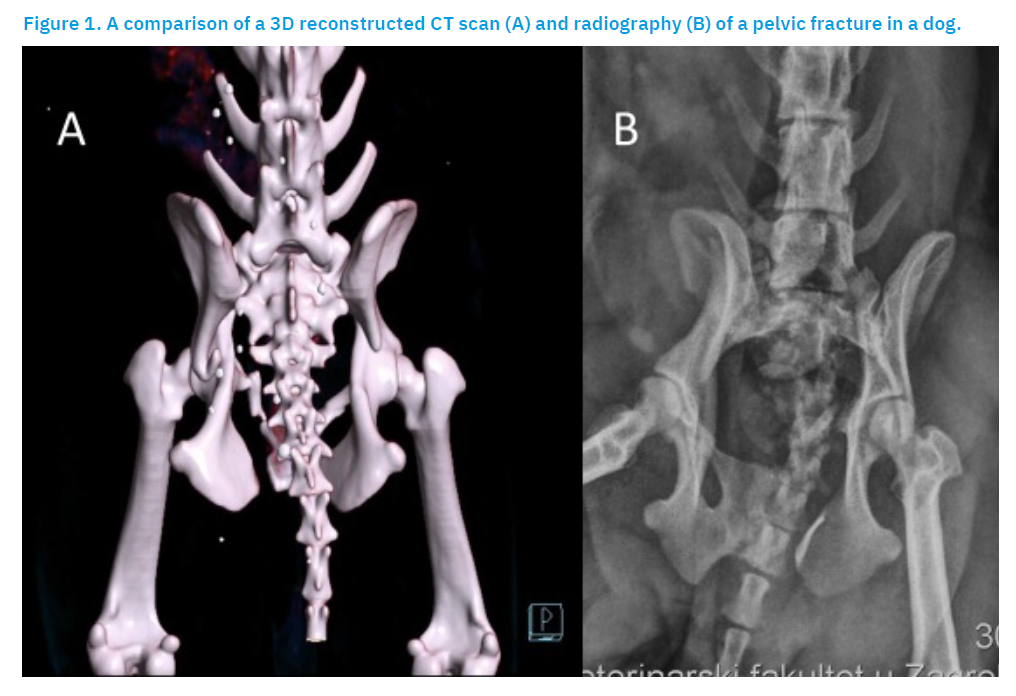
f) Computed tomography with three-dimensional reconstruction is more effective than conventional radiography for diagnosing and planning surgical management of certain pelvic fractures (Figure 1). CT is particularly useful for evaluating fragment configuration in complex acetabular fractures and for assessing the severity and location of fractures involving the sacrum (DeCamp et al., 2016). Though more accurate than radiography in the assessment of pelvic fractures (Lee et al., 2012), CT is significantly more expensive, increases radiation exposure, and is not available in all veterinary practices. CT is recommended in addition to radiography for sacral fractures with neurological signs, acetabular fractures, and when there is substantial uncertainty regarding fracture configuration. In most cases, radiography alone is sufficient for both diagnosis and treatment planning; however, CT imaging provides the surgeon with greater confidence in decision-making (Draffan et al., 2009).
Conservative management
Conservative management is indicated for fractures with minimal or no displacement, an intact acetabulum, and preserved pelvic ring continuity. The pelvic musculature provides natural stabilisation of fragments. Management consists of strict cage rest, restricted activity, and analgesic therapy. Bone healing typically occurs within 3–4 weeks (Remedios, 1999; DeCamp et al., 2016).
Surgical management
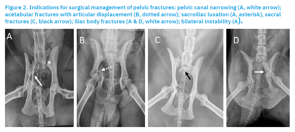
Surgical management is indicated in cases of (Figure 2):
- pelvic canal narrowing,
- acetabular fractures with articular displacement,
- sacroiliac luxation,
- sacral fractures,
- iliac body fractures,
- bilateral instability.
Due to associated soft tissue trauma, surgery may be delayed until stabilisation. However, optimal reduction is achieved within 3–5 days post-trauma. Fixation methods include Kirschner wires, external fixators, reconstruction plates, cortical screws, interfragmentary wires, or combinations thereof. Clinical experience indicates the highest success rates with reconstruction plates and screws.
Methods of surgical management
Sacroiliac Luxation/Fracture
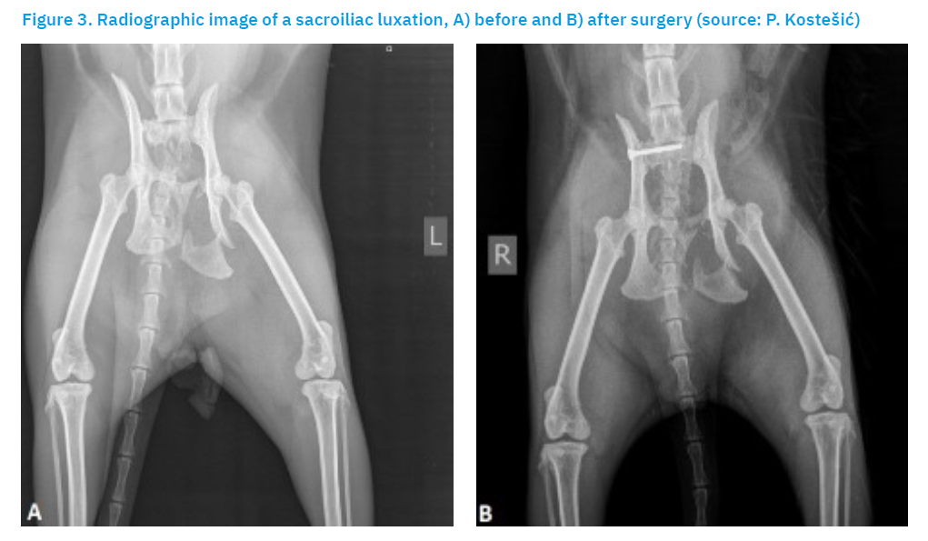
Sacroiliac (SI) luxation or fracture is a common injury following vehicular trauma, defined as traumatic separation of the ilial wing from the sacrum (Figure 3). Luxation resulting from blunt trauma is usually associated with additional soft tissue injuries, particularly within the pelvic cavity. Approximately 21% of all pelvic fractures are reportedly associated with sacroiliac luxation (Shales and Langley-Hobbs, 2005). In sacroiliac luxation, the ilium is typically displaced craniodorsally, and this displacement is invariably accompanied by fractures of the pubis and ischium or separation along the pelvic symphysis. Neurological injuries involving the lumbosacral trunk and/or sacral nerve roots may manifest as deficits in proprioceptive, voluntary motor, urinary, anal, and sensory reflexes. Fortunately, these deficits are reversible in most cases. Before surgical management, the function of the perineal, sciatic, and femoral nerves should be assessed (DeCamp et al., 2016). Surgical management may allow early weight-bearing on the affected limb and prevent narrowing or collapse of the pelvic canal (Tomlinson, 2012). Indications for internal fixation generally include pain, instability, and displacement compromising the pelvic canal or hip joint alignment. Contralateral pelvic limb injuries are common and may necessitate stabilisation of the SI joint to allow force distribution between the hind limbs and reduce stress on contralateral internal fixation (DeCamp et al., 2016).
Internal Fixation
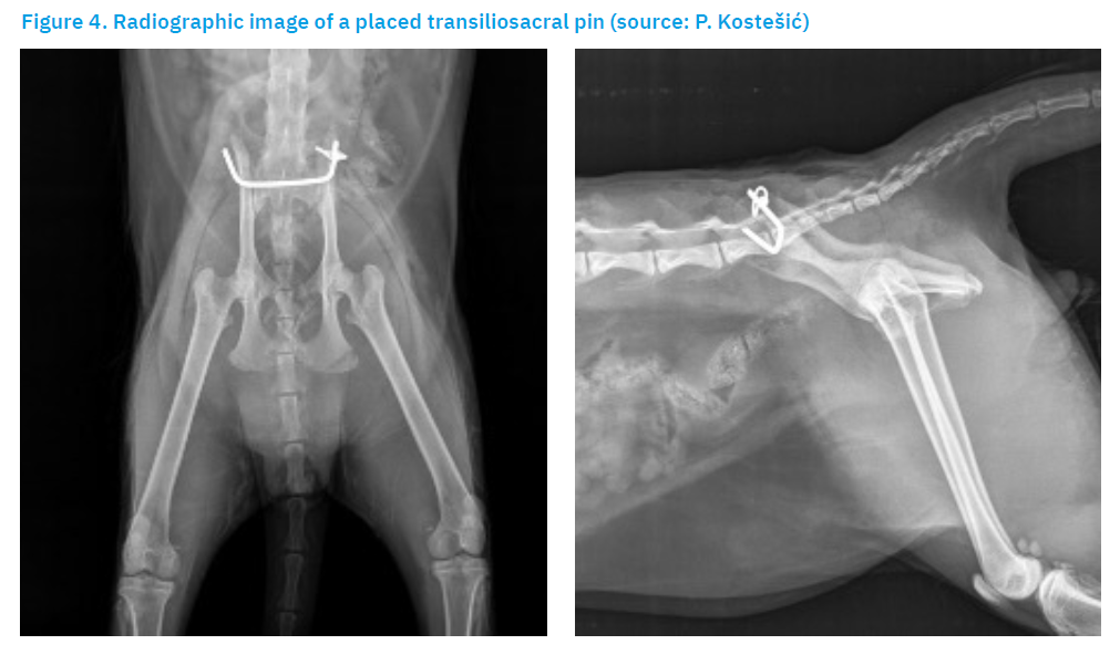
Stabilisation of the sacroiliac joint is achieved by inserting lag screws through the body of the ilium into the body of the sacrum. Two screws provide greater strength than a single screw of the same size; however, the size of the sacral body is a limiting factor in most dogs. Placement of two screws entirely within the sacral body is generally feasible only in very large dogs. In most dogs, it is essential to insert one long screw engaging approximately 60% of the sacral width. A glide hole should be drilled in the ilial body when fully threaded screws are used (DeCamp et al., 2016). Drilling the sacral body via a dorsal approach requires meticulous attention to anatomical detail (DeCamp et al., 2016). If the drill direction is excessively dorsal, there is a risk of penetrating the vertebral canal and damaging the cauda equina nerves. Minimising this risk is particularly important when using threaded implants, as nerve fibres may become entangled and irreversibly damaged. If the drill trajectory is too ventral, the screw may enter the pelvic canal, increasing the risk of screw loosening and haemorrhage from the medial sacral vessels (Shales and Langley-Hobbs, 2005). A second, shorter screw may be placed craniodorsal to the first. Screw length can be planned by measuring the distance on the dorsoventral radiographic projection. A second screw is desirable, particularly when part of the sacrum is fractured or when the first screw is not ideally positioned within the sacral body (DeCamp et al., 2016). However, one large, optimally positioned screw is preferable to two suboptimally placed screws (Shales and Langley-Hobbs, 2005). Implant loosening and subsequent loss of reduction remain the most common complications following screw fixation (Shales and Langley-Hobbs, 2005). Additional placement errors include screw insertion into lumbar articular processes, the lumbosacral disc space, or the seventh lumbar vertebra (DeCamp et al., 2016). Bilateral sacroiliac luxations or fractures account for approximately 23% of all SI luxations (Kaderly, 1991). Bilateral SI luxation may be stabilised via separate surgical approaches with lag screws placed on both sides (DeCamp et al., 2016), by a transilial pin, or by a transiliosacral pin (Parslow and Simpson, 2017).
This method uses a single transiliosacral pin designed to achieve bilateral reduction, with both ends shaped to compress the ilial wings. The pin is secured in a rotated position using either a screw or cerclage wire. The choice between cerclage wire and screws depends on the anatomy of the ilial wings and the final position of the rotated pin ends (Figure 4). Whenever possible, bilateral cerclage wires should be used, as this method is considered safer (Parslow and Simpson, 2017). This technique addresses several challenges associated with the reduction and treatment of these injuries by allowing direct bilateral visualisation, ensuring secure implant placement, and enabling precise reduction of SI luxation. Furthermore, it is a simple, safe, and cost-effective option for treating bilateral SI luxation. Long-term clinical outcomes are excellent. Transiliosacral fixation has recently been used with increasing frequency in small animal practice for stabilisation of bilateral SI luxations (Parslow and Simpson, 2017). Due to the limited space available for screw placement within the sacral body, screws inserted from opposite sides may contact or interfere with each other. Nevertheless, bilateral screw placement is almost always feasible (DeCamp et al., 2016).
Minimally Invasive Sacroiliac Fixation
The minimally invasive technique for sacroiliac fixation has proven to be an effective method for treating traumatic pelvic injuries in dogs. Preoperative pelvic radiographs in at least two projections are obtained to evaluate the fracture type and location, and to measure screw length. The total sacral width is measured along with the thickness of the ilium. A major drawback of this technique is the requirement for intraoperative fluoroscopy, which is costly and exposes personnel to radiation (Tomlinson, 2012). A minimally invasive technique for closed reduction and fixation of sacroiliac luxation in dogs has been described. A C-arm fluoroscope is used for imaging and placement of a Kirschner wire across the sacroiliac joint as temporary fixation (DeCamp et al., 2016). A 2012 study reported a screw loosening rate of 7% when the cumulative screw depth to sacral width ratio exceeded 60%, whereas a 48% loosening rate was observed when this ratio was less than 60% (Tomlinson, 2012). Although fluoroscopic guidance confirms initial drill guide orientation, successful screw placement ultimately depends on the surgeon’s ability to maintain optimal orientation during drilling. Even minor angular deviations from the ideal screw orientation perpendicular to the sagittal plane may have detrimental consequences (Déjardin et al., 2017). Advantages of this technique include minimal soft tissue damage, reduced postoperative pain and infection risk, accurate reduction, precise screw placement, low loosening rates, and early return to limb function on the affected side (Tomlinson, 2012). In addition to high equipment costs, radiation exposure remains a disadvantage; however, this exposure can be minimised by adhering to the ALARA (As Low As Reasonably Achievable) principle (Déjardin et al., 2017). The minimally invasive technique for repair of sacroiliac fractures or luxations is rapid, reliably more accurate, and less traumatic for patients compared with the open technique (Tomlinson, 2012).
Fracture of the Iliac Wing
As these fractures do not involve weight-bearing or articular regions, they are usually managed conservatively. In some cases, internal fixation may be performed for cosmetic reasons. Pins, interfragmentary wires, screws, or plates may be used (DeCamp et al., 2016).
Fracture of the Iliac Body
Fractures of the iliac body disrupt the weight-bearing axis of the pelvis and are therefore generally considered an indication for surgical management (Scrimgeour et al., 2017). Most iliac body fractures are oblique, with medial displacement of the caudal segment, resulting in narrowing or collapse of the pelvic canal. Some fractures are transverse or comminuted, and most are accompanied by fractures of the ischium and pubis. Neurological injuries of the lumbosacral trunk may accompany iliac fractures and should be evaluated prior to surgery. These injuries are often transient. Reduction and fixation of iliac body fractures help restore and stabilise fractures of the ischium and pubis. If the body of the ischium is also fractured, the hip joint will be unstable. The most common surgical management for iliac body fractures is internal fixation (DeCamp et al., 2016).
Open Approach and Reduction
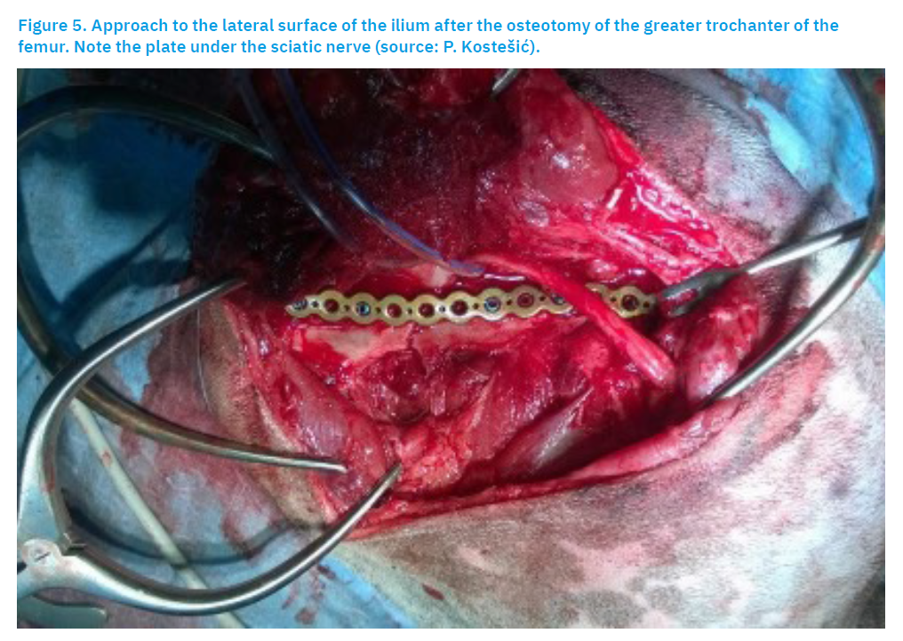
The lateral approach is the most commonly used technique for treating iliac body fractures, as it is considered the simplest and provides optimal visualisation of the fracture site (Scrimgeour et al., 2017) (Figure 5). The caudal segment should be elevated from beneath the cranial segment. Final reduction and fixation depend on the type of implant used (DeCamp et al., 2016).
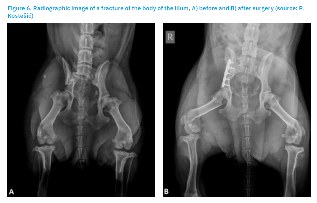
Fractures of the caudal portion of the iliac body are located immediately cranial and/or dorsal to the acetabulum, without involving the joint (Figure 6). If fixation is expected to extend over the dorsal aspect of the acetabulum, wider exposure can be achieved by performing an osteotomy of the greater trochanter of the femur (DeCamp et al., 2016).
Internal Fixation
Various methods for fixation of iliac fractures have been described and used. The highest success rates and ease of application are generally attributed to correct placement of a reconstruction plate on the lateral surface of the ilium. However, several authors have suggested ventral or dorsal plate placement to improve mechanical stability. A plate positioned on the ventral surface of the ilium is subjected to tensile forces, which provide the most favourable biomechanical environment for plate function, and may ultimately offer improved stability. Lateral fixation is technically straightforward and does not require an extensive inventory of implants. In selected cases, such as long oblique fractures and in relatively lean animals, insertion of lag screws or pins combined with tension band wiring may also be highly effective (DeCamp et al., 2016). A 2017 study described osteosynthesis using reconstruction T-plates via a standard lateral surgical approach to the ilium, with fracture reduction facilitated by bone-holding forceps applied to the greater trochanter or the ischial tuberosity (Scrimgeour et al., 2017).
Reconstruction Plates
The length and type of plate depend on the fracture location, with the limiting factor being the distance between the fracture line and the acetabulum. If sufficient space is available, a six-hole plate is recommended. Placement of one or more screws deeply into the sacral body significantly increases the anchorage strength of the cranial screws. The cranial portion of the iliac wing is thin and relatively soft, so screws may loosen easily in this region. Compression of the fracture line is desirable but rarely achievable due to the obliquity of most fractures. Two screws in the caudal segment are sufficient when stabilising a short bone fragment. If the caudal fragment is too short to accommodate two screws in a straight plate, T-plates or L-plates may be considered (DeCamp et al., 2016). The lateral surface of the ilium is concave, and plates used in this region generally require contouring (Guthrie and Kalff, 2017). The plate applied to the lateral surface should be contoured to a more pronounced concavity than the natural curvature of the ilium, as it tends to flatten postoperatively (DeCamp et al., 2016). The most common complication in the treatment of iliac body fractures with standard reconstruction plates is screw loosening. Screw loosening occurs in approximately 25% of cases, leading to loss of fracture reduction and narrowing of the pelvic canal (Scrimgeour et al., 2017).
Standard plates rely on friction between the plate and the underlying bone to achieve stability (Scrimgeour et al., 2017). Locking plates offer an advantage in thin bone by reducing the risk of screw loosening and subsequent loss of stability (Guthrie and Kalff, 2017).
Lag Screws
Studies have shown that two or more lag screws can provide effective stabilisation of oblique iliac body fractures when the fracture line length is at least twice the dorsoventral height of the ilium. Lesser obliquity does not allow screws to be placed at an appropriate angle to ensure angular stability (DeCamp et al., 2016).
Pins and Tension Band Wiring
Pins combined with tension band wiring may serve as an alternative to screw techniques, particularly in small breeds. At least two pins are required to provide angular stability, and the wire must be sufficiently strong to achieve interfragmentary compression. The wire may be placed around the protruding ends of the pins or between two short screws inserted into the ilium (DeCamp et al., 2016). The lateral approach to iliac fractures presents a mechanical challenge due to poor bone quality and difficulty in achieving a load-sharing construct. This may result in premature screw loosening and associated pelvic narrowing when standard plates are used (Scrimgeour et al., 2017).
Postoperative care
Following surgery, dogs are hospitalised for 24 to 48 hours, depending on the complexity of the procedure and the severity of concurrent conditions. During hospitalisation, routine clinical examinations are performed and neurological status is monitored (Paré et al., 2001; Guthrie and Kalff, 2017). When permitted, cold compresses may be applied to the surgical site for 15–20 minutes immediately after surgery to provide analgesia and reduce inflammation and oedema. The surgical wound should be monitored daily for drainage, discharge, excessive inflammation, or swelling. Patients should not be discharged with an actively draining or open wound. In patients with limb oedema, the affected limb should be elevated to prevent further swelling. This can be achieved by positioning the patient in lateral recumbency on the contralateral side and using a rolled towel or pillow to elevate the oedematous limb (Millis and Levine, 2014).
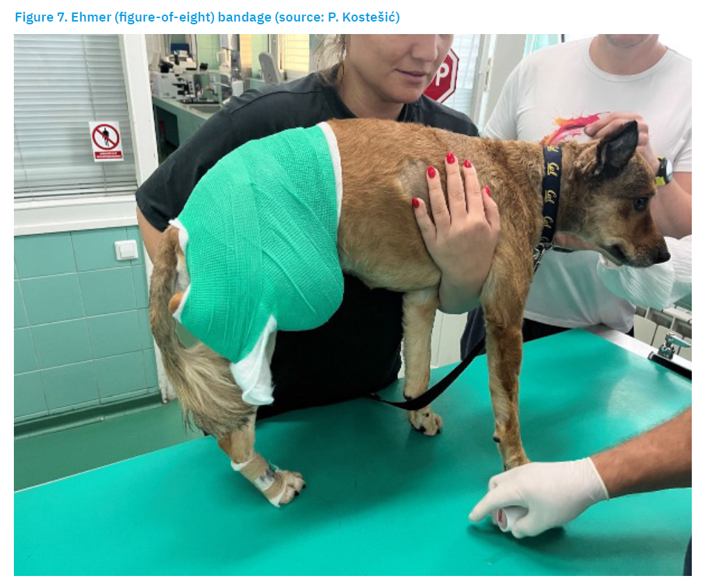
Warm compresses may be applied once the acute postoperative inflammatory phase has subsided, usually approximately 72 hours after surgery or injury. Application of heat increases local blood flow and may reduce muscle spasms (Millis and Levine, 2014). Soft tissue massage may help reduce postoperative oedema. The degree of limb oedema varies following orthopaedic surgery depending on patient size, procedure performed, and injury severity. Massage should begin distally and progress proximally to enhance lymphatic and venous return, thereby reducing oedema (Millis and Levine, 2014). Healing of pelvic fractures typically requires 6 to 10 weeks, which corresponds to the usual bone healing period. In general, reconstruction plates and screws are not removed unless clinically indicated (DeCamp et al., 2016). Muscles and tendons rapidly undergo atrophy during immobilisation or reduced activity, and other tissues such as cartilage, ligaments, and bone also undergo rapid changes in the postoperative period. Rehabilitation should begin as soon as the animal is stable, before complications associated with prolonged immobility develop (Millis and Levine, 2014). Pain management is a priority in orthopaedic surgery. Multimodal analgesia is now the standard of care for postoperative pain control in veterinary medicine (Millis and Levine, 2014). A transdermal fentanyl patch is an acceptable method of postoperative analgesia following complex orthopaedic procedures. Its ease of application and low cost make it an excellent option for postoperative pain management (Robinson et al., 1999). Additional medications commonly included in multimodal analgesic protocols are non-steroidal anti-inflammatory drugs (NSAIDs), opioids, and other analgesics (Millis and Levine, 2014). Activity restriction varies depending on the individual case, severity of trauma, and stability of fixation. In fractures involving the acetabulum, femoral head and neck, or in cases of mild hip luxation, local activity restriction may be achieved using an Ehmer sling, which may be maintained for up to two weeks (Millis and Levine, 2014; DeCamp et al., 2016) (Figure 7). According to the authors’ experience, maintaining an Ehmer sling for this duration is extremely uncomfortable for dogs and impractical in cats. The sling is designed to prevent weight-bearing on the affected limb and maintains the tarsus and hip in flexion with internal rotation of the hip (Millis and Levine, 2014). Schlag et al. (2019) found an increased risk, as well as a high incidence (50%) of soft tissue injury of any type associated with Ehmer sling application for craniodorsal hip luxations, and thus the recommendation for its use is considered guarded.
Physiotherapy may commence after several days and can be continued by the owner following discharge. Exercise restriction, consisting of leash walking only for elimination purposes, should be maintained for at least 6–8 weeks postoperatively. Gradual return to controlled exercise is recommended approximately 12 weeks after surgery (Piana et al., 2020). Follow-up radiographic examination is performed 6–8 weeks postoperatively (Wilson, 2015).
Postoperative patient care is an essential component of the rehabilitation programme. Successful outcomes depend on multiple factors, including the surgical management performed and owner expectations. Regardless of whether the patient has sustained an orthopaedic injury or undergone surgery, the ultimate goal remains the same: to support and facilitate functional recovery (Millis and Levine, 2014).
Discussion
Pelvic fractures are relatively common in dogs and cats. In most cases, they result from vehicular trauma and are often accompanied by additional injuries such as thoracic and abdominal trauma, as well as peripheral nerve damage. Although radiographic examination is generally sufficient for diagnosis, computed tomography is recommended when sacral or acetabular fractures are suspected. CT may alter the classification of a pelvic fracture; however, it rarely changes the therapeutic approach (Draffan et al., 2009). Treatment may be conservative or surgical. Conservative management is applied in approximately 75% of dogs with pelvic fractures in cases when fragment displacement is minimal or absent, the acetabulum is intact, and the continuity of the pelvic ring is preserved (DeCamp et al., 2016). Conservative treatment is based on analgesic therapy and activity restriction. Surgical management follows principles including anatomical reconstruction, stable fixation, and early mobilisation. Sacral fractures or sacroiliac luxation, fractures of the iliac body, and acetabular fractures generally require surgical management. Current experience indicates that the highest success rates in fracture reconstruction are achieved using reconstruction plates and screws. Postoperative care includes rest, activity restriction, and appropriate analgesic therapy, with follow-up radiographic examination performed 6–8 weeks postoperatively. Conservative management involves a prolonged recovery period and is associated with a higher risk of degenerative joint disease compared with surgical management. During conservative treatment, malunion or narrowing of the pelvic canal may occur. Furthermore, fracture malalignment or instability may lead to restricted hip joint motion, and improper alignment may result in persistent pain. Consequently, abnormal gait may develop. This treatment modality may not be adequate if restoration of normal biomechanical function is desired following therapy (Vassalo et al., 2015). Surgical management represents a more favourable prognostic option, as it enables earlier and improved patient recovery. However, degenerative joint disease may still develop if satisfactory anatomical reduction of fracture segments is not achieved, although minor deviations in reduction are generally considered acceptable. This review does not address acetabular fractures in detail, as their complexity and scope warrant a separate review. Publication of a second part of this paper focusing specifically on the treatment of acetabular fractures is planned.
Conclusions
Pelvic fractures account for at least one quarter of all fractures in dogs. Due to the rigid structure of the pelvis, considerable force (high-energy trauma) is required to produce a fracture; consequently, multiple fracture sites and injuries involving several organ systems are frequently observed. The most common cause of pelvic fractures is vehicular trauma. Pelvic fractures are often accompanied by concurrent injuries, including thoracic trauma, urinary tract injury, and peripheral nerve damage. Pelvic fractures are classified according to anatomical location as sacroiliac luxation or fracture, iliac wing fracture, iliac body fracture, acetabular fracture, ischial fracture, and fractures of the pelvic floor. Diagnosis is established by radiographic examination. Depending on the specific diagnosis, treatment may be conservative or surgical. Conservative management is employed in approximately 75% of cases and consists of cage rest and activity restriction.
Sacroiliac luxation, sacral fractures, acetabular fractures, and iliac fractures are the most common conditions requiring surgical management. Reconstruction plates and screws have demonstrated the highest success rates in fracture reduction and fixation. Postoperative management includes analgesia, activity restriction, physiotherapy, and follow-up radiographic evaluation to assess fracture healing. Surgical management of pelvic fractures in dogs represents a prognostically superior option, as it allows early mobilisation and is associated with a lower incidence of osteoarthritis compared with conservative management.
References [… show]
Prijelomi zdjelice u pasa: kirurške mogućnosti liječenja i postoperacijsko zbrinjavanje
Martina DRAŠIĆ1, drasicmartina@gmail.com; Niko IVKIĆ2, nivkic@vef.unizg.hr; Marko PEĆIN2, mpecin@vef.unizg.hr, orcid.org/0000-0002-1186-4774; Petar KOSTEŠIĆ2* (dopisni author), pkostesi@vef.unizg.hr, orcid.org/0000-0003-1603-7249; Siniša FARAGUNA3, sfaraguna@vef.unizg.hr, orcid.org/0009-0000-9397-5680.
1Veterinarska klinika Križevci d.o.o., 48260 Križevci, Hrvatska
2Klinika za kirurgiju, ortopediju i oftalmologiju, Veterinarski fakultet Sveučilišta u Zagrebu, 10000 Zagreb, Hrvatska
3Zavod za patofiziologiju, Veterinarski fakultet Sveučilišta u Zagrebu, 10000 Zagreb, Hrvatska
SAŽETAK
Cilj ovog preglednog rada bio je opisati najčešće postupke operacijskog liječenja lomova zdjelice u pasa. Procjenjuje se da lomovi zdjelice predstavljaju 25 % svih lomova u pasa. Lomovi zdjelice nastaju uslijed opsežne traume, a najčešće zbog udara automobila. Osim lomova, često su prisutne i druge ozljede poput traume prsnog koša, mokraćnog sustava te oštećenje perifernih živaca. S obzirom na anatomsku regiju, lomovi zdjelice dijele se na sakroilijačnu luksaciju/lom, lom krila crijevne kosti, lom tijela crijevne kosti, lom acetabuluma, lom sjedne kosti i lom zdjeličnog dna. Dijagnoza se temelji na RTG pretrazi. Liječenje lomova zdjelice može biti konzervativno ili kirurško. Većina lomova zdjelice liječi se konzervativno, što podrazumijeva restrikciju kretanja, odmor i određenu analgetsku terapiju. Oko 25 % lomova zahtijeva kirurško liječenje, a to su najčešće lomovi križne kosti ili sakroilijačna luksacija, lomovi tijela crijevne kosti i lomovi acetabuluma. Kirurško liječenje temelji se na redukciji i stabilizaciji loma. Od raznih implantata što se koriste prilikom kirurškog zahvata, najuspješnijima za željeni ishod pokazali su se vijci i rekonstrukcijske pločice. Za prihvatljiv ishod operacijskog zahvata neophodna je savršena anatomska pozicija, stabilna fiksacija i rana pokretljivost. Poslijeoperacijska skrb obuhvaća analgetsku terapiju, ograničenje aktivnosti i kontrolnu RTG pretragu u roku 6–8 tjedana nakon operacije.
Ključne riječi: lomovi zdjelice; pas; vijci; rekonstrukcijske pločice; analgetska terapija; RTG.
