
High-mortality outbreak of Yersinia pseudotuberculosis infection in a guinea pig herd: a case report
D.Novosel, G. Kompes, B. Habrun and A. Jungić*
Dinko NOVOSEL 1*, novosel@veinst.hr, orcid.org/0000-0003-2602-8696; Gordan KOMPES 2, kompes@veinst.hr, orcid.org/0009-0000-4934-1357; Boris HABRUN 2, habrun@veinst.hr, orcid.org/0009-0002-9688-026X; Andreja JUNGIĆ 3 (corresponding author), jungic@veinst.hr, orcid.org/ 0000-0002-9497-9904.
1Laboratory for Pathology, Department of Pathological Morphology, Croatian Veterinary Institute, 10000 Zagreb, Croatia
2Laboratory for General Bacteriology and Mycology, Department of Bacteriology and Parasitology, Croatian Veterinary Institute, 10000 Zagreb, Croatia
3Laboratory for Rabies and General Virology, Department of Virology, Croatian Veterinary Institute, 10000 Zagreb, Croatia

https://doi.org/10.46419/cvj.57.3.2
Abstract
Yersinia pseudotuberculosis caused a fulminant outbreak in a Croatian breeding colony of 84 guinea pigs, in which 80 animals (95%) died within 72 hours after abrupt food refusal and marked lethargy, without diarrhoea. Seven freshly dead animals were examined. At necropsy, 2–5 mm, white-yellow nodules were found distributed in the liver, spleen, lungs, mesenteric lymph nodes and partly also in the small intestine. Aerobic culture of these organs on Columbia blood agar (28°C, 24–48 h) revealed pure growth of Y. pseudotuberculosis. Histology showed well demarcated necro-pyogranulomatous lesions with central bacterial colonies, margins of neutrophils and a peripheral coat of macrophages and few lymphocytes. Immunohistochemistry confirmed abundant lysozyme-positive macrophages and few CD3-positive T and CD79α-positive B cells, indicating a predominantly innate response. This case report emphasises the extreme lethality and rapid progression of guinea pig yersiniosis and highlights its zoonotic potential.
Key words: Yersinia pseudotuberculosis; guinea pig; histopathology; immunohistochemistry
Introduction
Yersinia pseudotuberculosis, a small, Gram-negative, and facultatively anaerobic pleomorphic coccobacillus, is the causative agent of clinical yersiniosis. This bacterium has been classified into 21 serotypes based on the O antigen, whereby only some serotypes are considered pathogenic. Serotype O:1, most common in Europe, has been isolated from wild boar, rodents and birds and detected in infections in zoological facilities (Tsubokura et al., 1989; Galosi et al., 2015; Arrausi-Subiza et al., 2016; Le Guern et al., 2016; Hammerl et al., 2021). Outside Europe, other serotypes have been associated with disease in wild animals and in zoos (Nakamura et al., 2016). For example, serotype 3 has been isolated from various deer species in the United States (Sanford, 1995), and serotypes 1b, 2b, 3, 4b, 6 and 7 have been found in deceased monkeys of various species in Japan (Iwata et al., 2008; Nakamura et al., 2009).
In addition, avirulent strains of Y. pseudotuberculosis are known to occur in wild animals and in the environment (Nagano et al., 1997), emphasising its wide distribution and the possibility that most animals are asymptomatic carriers. This pathogen poses a serious threat to guinea pigs, rabbits, cats, birds and non-human primates in laboratories, where stress and low temperatures can trigger symptomatic outbreaks of otherwise asymptomatic infections, and it is also a significant concern due to its wide host range and persistence in the environment. Due to their ability to cause disease under stress or at low temperatures, infection can rapidly progress to become fatal. Interestingly, certain species such as the white rat, hamster and lower vertebrates such as fish, amphibians and reptiles are resistant to this infection.
The disease has been transmitted primarily via the faecal-oral route and has been linked to enteric yersiniosis, which affects humans as well as domestic and wild mammals, and causes symptoms such as lymphadenitis of the mesenteric lymph nodes, terminal ileitis and appendicitis (Galosi et al., 2015; Arrausi-Subiza et al., 2016; Le Guern et al., 2016; Hammerl et al., 2021). Infection with Y. pseudotuberculosis in animals often leads to a chronic form of the disease in which the animals show symptoms such as weight loss and diarrhoea, leading to death within three to four weeks. In more acute cases, the disease can lead to rapid death with visible miliary changes in the liver and spleen (Nakamura et al., 2016). Interestingly, Y. pseudotuberculosis is also known to cause “Far East scarlet-like fever” in humans, particularly in Asia, demonstrating its zoonotic potential and ability to cause various clinical manifestations in humans (Sanford, 1995). Its ability to survive in the environment at low temperatures (4 to 20°C) increases the risk of epizootics, necessitating continuous monitoring and preventive measures in zoological collections and in the general population (Sanford, 1995). Overall, Y. pseudotuberculosis represents a significant pathogen for both wildlife and domestic animals that requires thorough attention and an interdisciplinary approach to research, diagnosis and control in order to minimise the health risks to animals and humans. Y. pseudotuberculosis is primarily transmitted via the faecal–oral route through the consumption of food or water contaminated with faeces from natural reservoirs such as birds and rodents (Le Guern et al., 2016; Nakamura et al., 2016; Hahn et al., 2021).
Yersiniosis has a high mortality rate, which is often only detected post-mortem, with underreported cases possibly distorting mortality rates (Nederlof et al., 2025). In mammals, it manifests primarily as a gastrointestinal disease characterised by ulcerative enteritis, mesenteric lymphadenomegaly and hepatitis, while in birds it is described as a lymphoreticular disease, mainly manifesting as hepatitis and splenitis (Cork et al., 1999). Symptoms in mammals typically include anorexia, lethargy and sometimes diarrhoea, with sudden death occurring without prior signs in various species including scimitar-horned oryx and Seba’s short-tailed bats (Nederlof et al., 2025). Neurological symptoms such as paraplegia and ataxia have also been observed (Hammerl et al., 2021). Cervids often succumb suddenly and show symptoms such as diarrhoea and emaciation before death (Sanford, 1995; Ceccolini et al., 2020). Yersiniosis in birds often results in sudden death with symptomatic lethargy and diarrhoea preceding death, with notable cases occurring in blue-fronted and yellow-headed amazons exhibiting bright green faeces (Galosi et al., 2015). In non-human primates and other mammals, yersiniosis can lead to systemic disease with lethal outcome, often manifesting in a subtle manner, with symptoms developing rapidly before death (Buhles et al., 1981; Mingrone and Fantasia, 1988; Bielli et al., 1999; Krylova and Dzhikidze, 2000; Kageyama et al., 2002; Nakamura et al., 2009; Iwata and Hayashidini, 2011; Zao et al., 2013; Soto et al., 2015; Zhao et al., 2016; Walker et al., 2018; Ceccolini et al., 2020; Hammerl et al., 2021).
The presence of the ypmA gene has been associated with atypical disease manifestations such as skin rash and arthritis in primates (Nakamura et al., 2009). At necropsy, multifocal white-yellow nodules are frequently found in the liver, spleen, lungs and mesenteric lymph nodes, occasionally also in the intestine and kidneys (Nederlof et al., 2025). These nodules may be present without causing visible tissue changes (Owston et al., 2006). Respiratory lesions often include white-yellow pulmonary nodules, bronchopneumonia and signs of pulmonary problems such as hyperaemia, oedema and petechiae, with acute fibrinopurulent or necro-suppurative pneumonia occurring in severe cases (Gombač et al., 2008; Hahn et al., 2021; Cano-Terriza et al., 2022). Gastrointestinal disorders are observed, in particular haemorrhagic enteritis, which mainly affects the small intestine and less frequently the stomach and colon (Ceccolini et al., 2020). The spleen and liver often show multifocal necrotising splenitis and hepatitis, with these organs being enlarged and nodular, but rarely hyperaemic (Nederlof et al., 2025). Renal and cardiac manifestations are less common but may include renal hyperaemia and petechiae, as well as cardiac petechiae and ecchymoses (Nakamura et al., 2015; Womble et al., 2022). A unique case of adrenal inflammation in a paca and skin petechiae in silver monkeys illustrated the different manifestations of this infection in different species (Fogelson et al., 2015; Ceccolini et al., 2020). These findings emphasise the ability of Y. pseudotuberculosis to cause extensive and diverse pathological changes affecting multiple organ systems in various mammalian and avian taxa. Histopathological changes associated with Y. pseudotuberculosis infection include multifocal necrosis in the liver and spleen, characterised by central bacterial colonies surrounded by necrotic cellular debris, vacuolated macrophages and neutrophils. Hypereosinophilic necrotic hepatocytes or splenic tissue is found at the edges of the lesions (Fogelson et al., 2015). This pattern of coagulative and lytic hepatic necrosis with neutrophilic inflammation is common in various taxa, including birds (Ceccolini et al., 2020). The intestines often show microabscesses in the lamina propria and severe lesions around the Peyer’s patches, characterised by mucosal ulceration and haemorrhage.
Histological examination shows densely packed bacterial colonies in necrotic mucosal areas, accompanied by fibrin, neutrophils, macrophages and lymphocytes extending into the submucosa (Nakamura et al., 2009; Fogelson et al., 2015). Aberrant bacterial morphologies, such as spherical or filamentous forms, may mimic fungi or protozoa and require specific immunohistochemical staining (IHC) for accurate identification (Nakamura et al., 2015; Womble et al., 2022). In severe cases, myocardial degeneration and pulmonary oedema are observed, particularly in squirrel monkeys, while the mesenteric lymph nodes show oedema with significant neutrophil and macrophage infiltration (Nakamura et al., 2009). Far East Scarlet-like Fever (FESLF), also known as Izumi fever in Japan, is a severe inflammatory disease that was first described in 1959 during an epidemic in Vladivostok, Russia, in which over 300 patients were hospitalised. Since then, several epidemics and sporadic cases have occurred in Russia and Japan, often linked to the consumption of contaminated food. FESLF is characterised by symptoms such as skin rash, hyperaemia of the tongue and peeling of the skin.
The causative agent, Yersinia pseudotuberculosis, usually causes self-limiting gastroenteritis in Europe, but Far Eastern strains can produce a superantigenic toxin, Y. pseudotuberculosis-derived mitogen A (YPMa), which leads to more severe symptoms. Yersinia pseudotuberculosis was first isolated in 1883 and is known for its pathogenicity, particularly its ability to multiply in cold environments as low as 4°C, allowing it to contaminate food. The disease is so significant that it has been included in the national health reporting systems in Russia and Japan since 1988. Epidemiological studies show that the pathogen has a broad reservoir in animals, leading to disease on all continents. The difference in clinical presentation and severity between Europe and Asia is due to the virulence factors of the strains from these regions, with the production of YPMa by the Far Eastern strains being particularly striking. The epidemiology of FESLF has evolved. It was originally confined to the Russian Far East between 1959 and 1980, and later spread throughout Russia due to socio-economic changes (Tseneva et al., 2012). Today, FESLF has been recognised as a national health problem in Russia. A significant number of new cases occur each year, mainly affecting children, with seasonal peaks in the colder months due to local agricultural practises. Clinically, FESLF begins with symptoms similar to those of scarlet fever, but rapidly evolves into a more complex gastrointestinal and systemic inflammation. This necessitates increased public health surveillance and preventive strategies during peak transmission periods. Knowledge of the link between FESLF and YPMa has spurred targeted research and public health initiatives to mitigate the impact of the disease. This emphasises the need for ongoing research and adaptive public health measures to effectively manage and control this geographically widespread disease.
Material and Methods
After the death of approximately 80 animals, of which only four survived, seven diseased guinea pigs were referred to the pathology laboratory of the Croatian Veterinary Institute. At the beginning of the outbreak, the herd owner observed that the affected animals suddenly stopped eating and became clearly lethargic; no diarrhoea or other obvious signs of enteritis were observed. Within 72 hours, seven guinea pigs died suddenly and all seven were necropsied. Gross pathoanatomical changes were described and photo-documented, and tissue samples were fixed in 10% buffered formalin for histopathology. Portions of the liver, spleen, lung, mesenteric lymph nodes, and small intestine were aseptically removed and sent to the Department of Bacteriology for culture. Samples were immediately inoculated onto Columbia blood agar plates supplemented with 5% defibrinated sheep blood and incubated aerobically at 28°C for 24–48 hours. After incubation, colonies with a small, round, smooth and greyish-white morphology were selected for further analysis. Gram staining was performed on the suspect colonies, which revealed Gram-negative, rod-shaped bacteria. Biochemical characterisation was performed using standard laboratory tests including catalase, oxidase, urease, indole production, citrate utilisation and fermentation of glucose, lactose, sucrose, mannitol and sorbitol. Organs were examined histopathologically for microorganisms using standard Mayer’s H&E staining technique and Brown and Brenn staining.
For the detection of CD3, CD79a and lysozyme, tissue sections were deparaffinised in xylene and rehydrated through graded alcohols. Endogenous peroxidase activity was blocked by incubating the sections with 3% hydrogen peroxide in 0.1 M Tris-buffered saline (TBS, pH 7.6) for 30 minutes. IHC was performed with three primary antibodies: Anti-Human-CD3, Anti-CD79a, and Anti-Lysozyme, with the specific pretreatment protocols listed in Table 1.
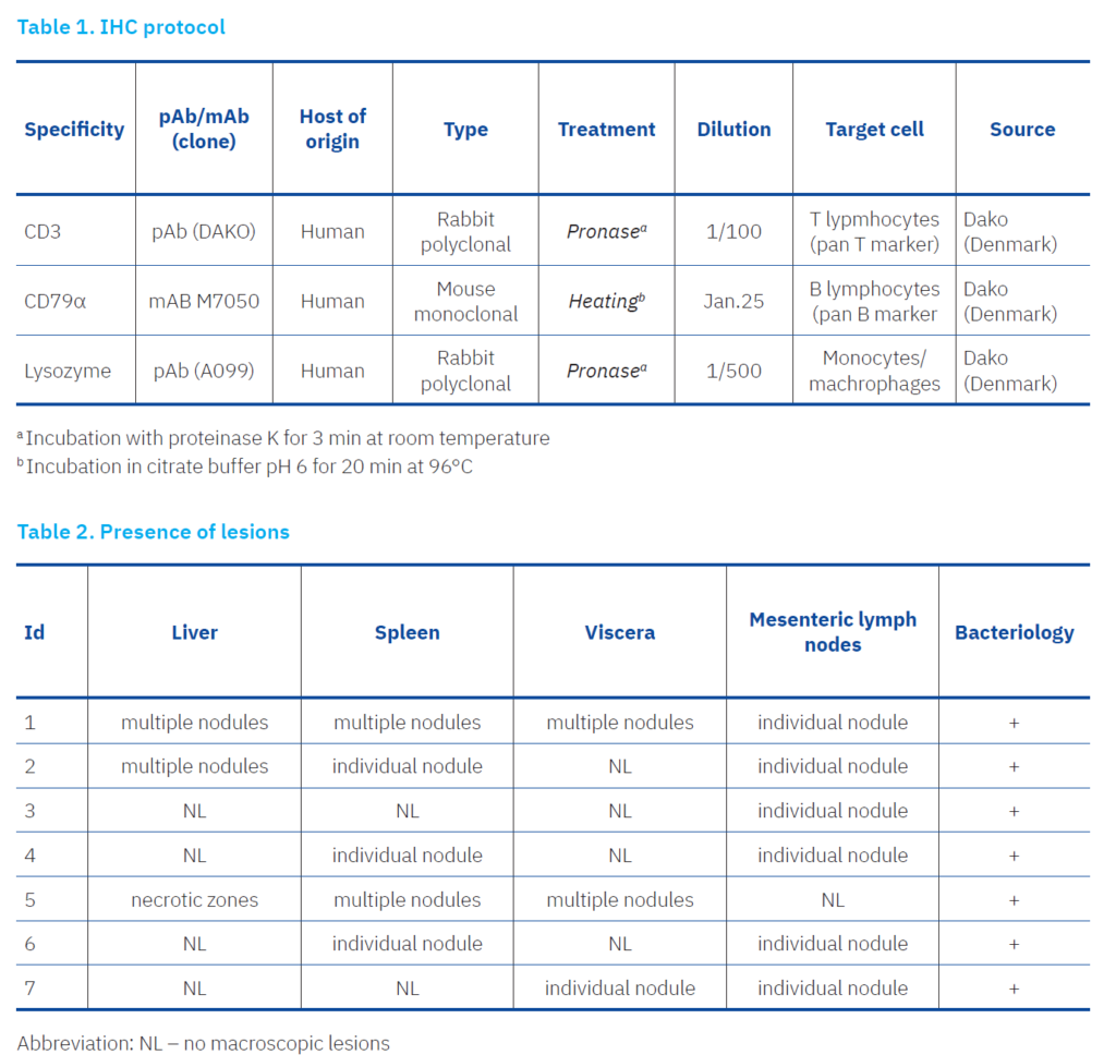
Visualisation of positive reactions was performed using an HRP-conjugated anti-rabbit and anti-mouse polymer detection system (Envision, Dako), followed by a 5-minute incubation in diaminobenzidine (DAB) hydrogen peroxide solution (Dako, Denmark). Slides were counterstained with Mayer’s haematoxylin, dehydrated, covered with coverslips and examined microscopically. The negative controls contained irrelevant primary antibodies at the same dilution. The IHC micrographs were digitally processed using QuPath software. The positive IHC signal, represented by the DAB chromogen, was annotated and visualised with the red RGB colour 204, 51, 102.
Results
The identity of isolates as Y. pseudotuberculosis was confirmed based on colony morphology, Gram staining characteristics and biochemical test profiles consistent with the species using standard identification protocols. The colonies had a small, round, smooth and grey-white morphology and the isolates were catalase-positive, oxidase-negative, urease-positive and able to ferment glucose without gas formation, but negative for lactose and sucrose fermentation. Necropsy revealed identical pathoanatomical changes in all seven guinea pigs examined (Table 2).
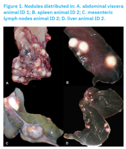
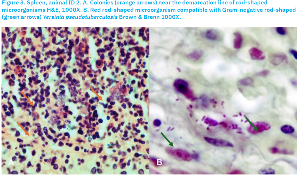
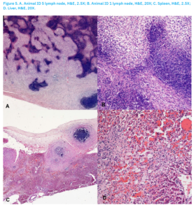
Disseminated nodules with a diameter of 2 to 5 mm were described in the lungs, liver, spleen, visceral lymph nodes and abdominal viscera (Figure 1). In cross-section, the nodules appeared as dense white-yellowish cheesy masses. These nodules were sharply demarcated and protruded from the surrounding tissue. Histopathological examination revealed necrotising granulomatous changes in the parenchymal organs, lymph nodes and abdominal viscera. There was a zone of caseous necrosis in the centre, which merged into a purulent-necrotic mass towards the edge. Along the margin was a belt of mixed polymorphonuclear cells, macrophages and a small number of lymphocytes, merging into a zone of proliferative cells (Figure 2). The H&E staining technique showed colonies with some bacteria in the necrotic area (Figure 3a). In Brown & Brenn staining, rod-shaped (Figure 3b), red-coloured microorganisms were observed.
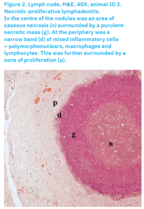
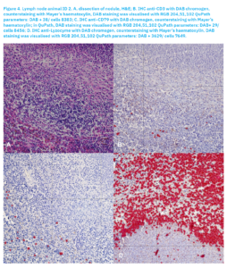
Histopathological and immunohistochemical examination showed that the nodules had a characteristic structure. The nodule was primarily encapsulated and undoubtedly represents a granulomatous-pyonecrotic lesion. The central part was a zone of necrosis mixed with neutrophils towards the outside, followed by a capsule that showed a strong mononuclear infiltration both inside and outside, consisting predominantly of histiocytes/macrophages, rarely with T or B lymphocytes. In the zone where T or B lymphocytes were present, macrophages contained a small amount of haemosiderin in the cytoplasm.
In addition to the nodules in the mesentery, the most significant lesions were found in the liver, spleen, and lymph nodes. While the nodules in the liver and spleen were clearly visible macroscopically, the changes in the lymph nodes were microscopic. In addition to the nodules, which had a characteristic structure as already described, atrophy and fatty degeneration of the hepatocytes were observed in parts of the liver. In most areas, the normal histological architecture of the liver was no longer recognisable and hepatocytes were not present. There was severe mononuclear infiltration, some distortion in the sinusoids and vasculitis. The lymph nodes also lacked the typical architecture, lymphoid follicles were not recognisable and most areas were dominated by a purulent-necrotic mass with considerable histiocytic proliferation.
Discussion
It was not unexpected that Y. pseudotuberculosis was confirmed to circulate in Croatia, since previous unpublished reports suggested its presence as a pathogen, and its ability to cause gastrointestinal problems in humans. However, no highly pathogenic strains have been reported circulating since FESLF, or at least not in significant numbers. It was also known that the disease had considerable zoonotic potential and presents a risk.
In the present study, the samples show well-defined nodules with a central zone of caseous necrosis surrounded by a purulent-necrotic mass and a narrow band of mixed inflammatory cells (polymorphonuclear cells, macrophages and lymphocytes), as previously described (Owston et al., 2006; Fogelson et al., 2015). However, in contrast to some previous reports, no marked effusion was observed in the pleural or peritoneal cavity, which could indicate differences in the clinical presentation of the disease or the stage of pathological changes. The inflammatory reaction observed was mixed. Neutrophils dominated the central part of nodules, while the surrounding area was predominantly occupied by macrophages, as the IHC showed mainly these cells. There were very few lymphocytes, and when present, T lymphocytes were slightly more abundant than B lymphocytes. This was indicative of the nature of the immune response, which appears to be primarily non-specific, while any specific immune response was more likely to be cellular than humoral. There is certainly room for further research, primarily to perform genotyping of the circulating strains to assess the risk to humans and to investigate the immune response in more detail, specifically which T lymphocytes are involved.
References [… show]
Izbijanje infekcije bakterijom Yersinia pseudotuberculosis s visokim mortalitetom u uzgoju zamorčića – prikaz slučaja
Dinko NOVOSEL 1*(dopisni autor), novosel@veinst.hr, orcid.org/0000-0003-2602-8696; Gordan KOMPES 2, kompes@veinst.hr, orcid.org/0009-0000-4934-1357; Boris HABRUN2, habrun@veinst.hr, orcid.org/0009-0002-9688-026X; Andreja JUNGIĆ3(dopisni autor), jungic@veinst.hr, orcid.org/ 0000-0002-9497-9904.
1Laboratorij za patologiju, Odjel za patološku morfologiju, Hrvatski veterinarski institut, 10000 Zagreb, Hrvatska
2Laboratorij za opću bakteriologiju i mikologiju, Odjel za bakteriologiju i parazitologiju, Hrvatski veterinarski institut, 10000 Zagreb, Hrvatska
3Laboratorij za bjesnoću i opću virologiju, Odjel za virologiju, Hrvatski veterinarski institut, 10000 Zagreb, Hrvatska
Yersinia pseudotuberculosis izazvala je fulminantnu epizootiju u hrvatskoj uzgojnoj koloniji od 84 zamorčića od kojih je 80 (95 %) uginulo u roku od 72 sata. Prije uginuća, zamorčići su pokazivali znakove izražene letargije, odbijali su hranu, no bez prisutnog proljeva. Obdukciji je podvrgnuto sedam svježe uginulih jedinki. Pri nekropsiji su zapaženi 2–5 mm veliki, bijelo-žuti čvorići rasprostranjeni u jetri, slezeni, plućima, mezenterijalnim limfnim čvorovima te djelomično u tankom crijevu. Aerobnom kultivacijom tih organa na krvnom agaru Columbia (28 °C, 24–48 h) u svih je životinja dokazan rast Y. pseudotuberculosis. Histološki su utvrđene dobro ograničene nekro-piogranulomatozne lezije s centralnim bakterijskim kolonijama, rubnim slojem neutrofila i perifernim plaštom makrofaga uz malobrojne limfocite. Imunohistokemijskom metodom utvrđena je obilna prisutnost lizozim-pozitivnih makrofaga te rijetkih CD3-pozitivnih T- i CD79α-pozitivnih B-stanica, što uglavnom upućuje na urođeni imunološki odgovor. Ovaj prikaz slučaja naglašava iznimnu letalnost i brzi tijek jersinioze u zamorčića te ističe njezin zoonotski potencijal.
Ključne riječi: Yersinia pseudotuberculosis, zamorčić, histopatologija, imunohistokemija
